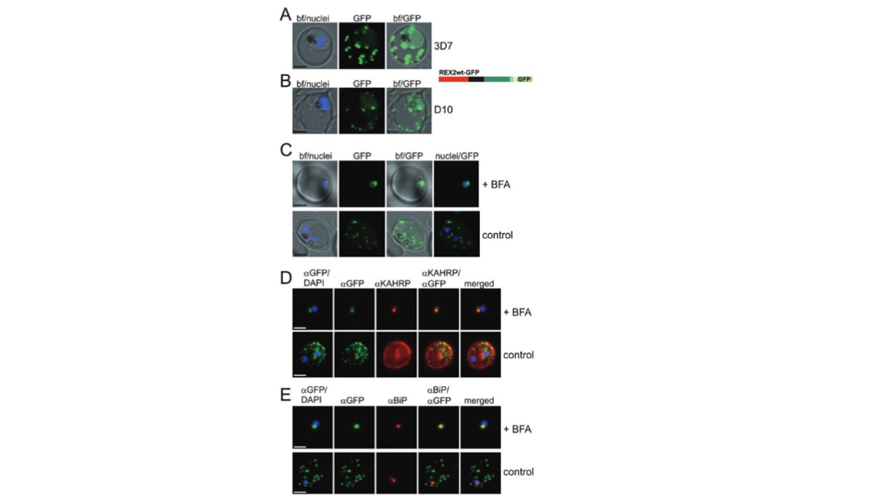
Export of REX2GFP does not depend on genes on the right arm of chromosome 9 and is sensitive to BFA.A and B. GFP fluorescence in live 3D7 (A) or D10 (B) parasites expressing wild-type REX2GFP from an episomal vector. GFP fluorescence appears as dots in the host cell cytoplasm typical for Maurer’s cleft localization. Nuclei are stained with DAPI(blue); merges with bright field images (bf) are shown. A schematic structure of the episomally expressed REX2 is shown beside the panel: red, N-terminus; black, TM; dark green, C-terminus; light green, GFP.C. Live cell fluorescence of 3D7 parasites expressing REX2GFP treated with BFA. Panels are shown as in (A) and (B) except for additional merges between nuclei and GFP(nuclei/GFP). Treated cells (+BFA) and mock-treated cells (control) are shown. D. IFA of acetone-fixed infected RBCs expressing REX2GFP and stained with anti-GFP (green) and anti-KAHRP (red) antibodies. Overlays are shown as indicated above each panel. The last panel shows an overlay of all three images (merged). E. IFA of acetone-fixed infected RBCs expressing REX2GFP and stained with anti-GFP (green) and anti-BiP (red) antibodies. Overlays of panels are shown as in (D). Size bars are 2 mm. Haase S, Herrmann S, Grüring C, Heiber A, Jansen PW, Langer C, Treeck M, Cabrera A, Bruns C, Struck NS, Kono M, Engelberg K, Ruch U, Stunnenberg HG, Gilberger TW, Spielmann T. Sequence requirements for the export of the Plasmodium falciparum Maurer's clefts protein REX2. Mol Microbiol. 2009
Other associated proteins
| PFID | Formal Annotation |
|---|---|
| PF3D7_0202000 | knob-associated histidine-rich protein |
| PF3D7_0936000 | ring-exported protein 2 |