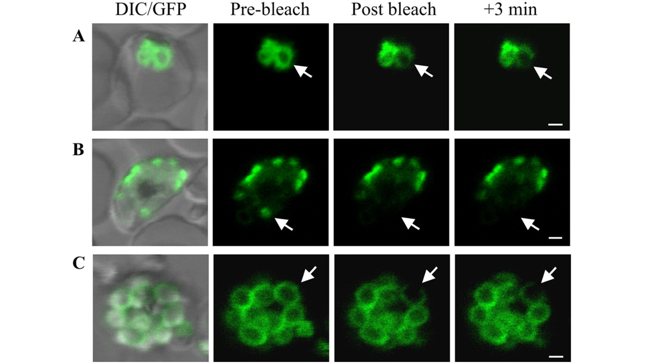
Photobleach analysis of PfGAP50-GFP organization. The panels are prebleach images with and without DIC overlay and postbleach images after application of a laser pulse at the positions indicated by arrows. (A) A region of the ER in an early-trophozoite stage parasite shows complete loss of fluorescence at the point of bleaching, partial loss from a connected compartment, and little loss from an adjacent compartment. There was no recovery after 3 min. (B) Application of a laser pulse to PfGAP50-GFP in an apical cap in an early schizont ablates fluorescence with no recovery from adjacent structures. (C) Spot bleaching of a region of PfGAP50-GFP in a region of a mature merozoite results in complete loss of fluorescence at the point of bleaching, with little loss from other regions of the same merozoite or from adjacent merozoites. Bars = 1 μm.
Yeoman JA, Hanssen E, Maier AG, Klonis N, Maco B, Baum J, Turnbull L, Whitchurch CB, Dixon MW, Tilley L. Tracking Glideosome-associated protein 50 reveals the development and organization of the inner membrane complex of Plasmodium falciparum. Eukaryot Cell. 2011 10(4):556-64.