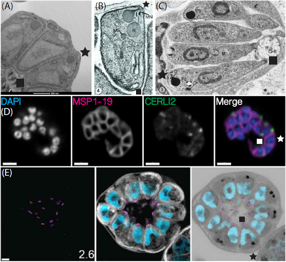
Images of fixed malaria schizonts demonstrating how the merozoites are often organised in a wedge pattern around the residual body with their wider ends facing out of the cell. (A–C) Electron micrographs of a Plasmodium falciparum schizont (A), an individual Plasmodium knowlesi merozoite inside a schizont (B), and a Plasmodium elongatum schizont (C). (D) Immunofluorescence microscopy images of a P. falciparum schizont stained with DAPI (4′,6-diamidino-2-phenylindole, nucleus), an anti-MSP1-19 antibody (merozoite surface), and an anti-HA (hemagglutinin) antibody (detects HA-tagged rhoptry protein CERLI2. (E) Expansion microscopy images of a P. falciparum schizont stained
with an anti-V5 antibody (basal complex, magenta), NHS ester (N-Hydroxysuccinimide, protein density stain, greyscale), BODIPY TRc (lipids, white), and SYTOX (nucleus, cyan). These images clearly show that the nuclei of the merozoites are located at the narrower end (indicated by a square) in closest proximity to the residual body while the rhoptries are positioned at the wider end (indicated by a star) facing out of the cell. Andrews M, Baum J, Gilson PR, Wilson DW. Bottoms up! Malaria parasite invasion the right way around. Trends Parasitol. 2023 39(12):1004-1013.
Other associated proteins
| PFID | Formal Annotation |
|---|---|
| PF3D7_0405200 | cytosolically exposed rhoptry leaflet interacting protein 2 |