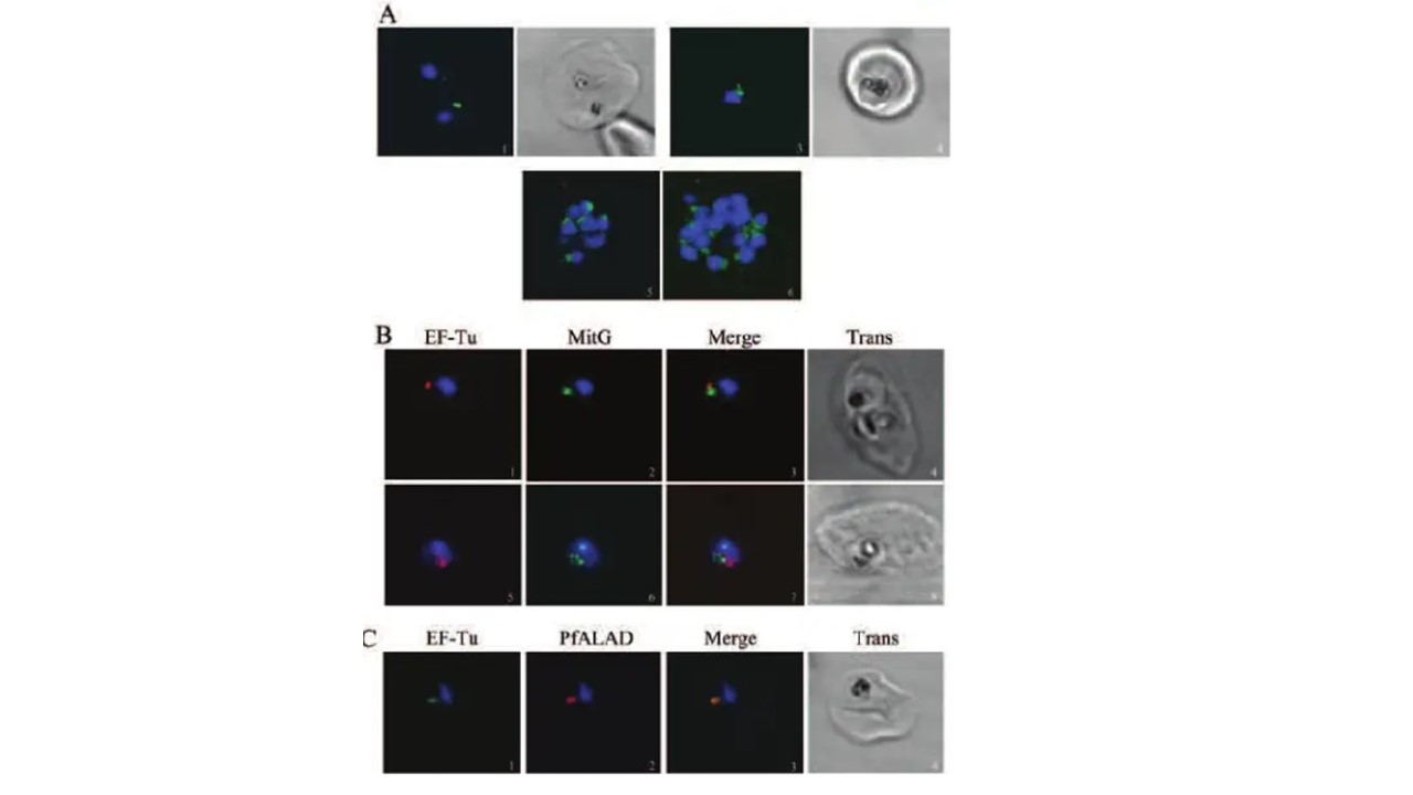
EF-Tu is localized within the apicoplast of P. falciparum. A. Fluorescence (green) localizes within the apicoplast in the middle trophozoite (1), late trophozoite (3) and schizont (5 and 6) stages. Images 2 and 4 are the corresponding phase contrast scans of 1 and 3 respectively. Green fluorescence is associated with most nuclei(blue) in images 5 and 6 (all apicoplasts may not lie in the photographed focal plane). B. Closely associated but distinct fluorescence for apicoplast (red) and mitochondria (green) is seen in the middle trophozoite (1–4) and early schizont stages (5–8).C. Overlapping signals are observed for EF-Tu (green) and PfALAD (red) in the middle trophozoite stage. Chaubey S, Kumar A, Singh D, Habib S. The apicoplast of Plasmodium falciparum is translationally active. Mol Microbiol. 2005 56(1):81-9. PMID: 15773980.
Other associated proteins
| PFID | Formal Annotation |
|---|---|
| PF3D7_1246100 | delta-aminolevulinic acid synthetase |
| PF3D7_1330600 | elongation factor Tu, putative |
| PF3D7_1348300 | elongation factor Tu, putative |
