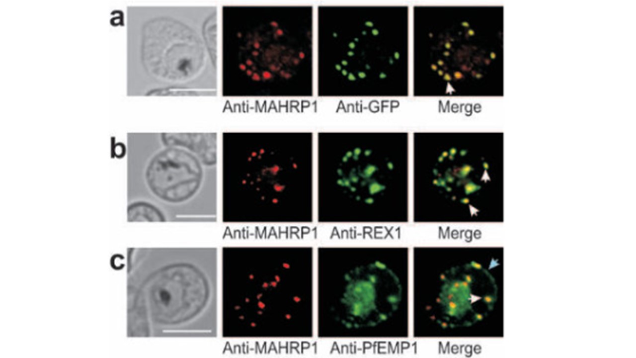
Immunofluorescence microscopy of MAHRP11-249 -GFP.
(a) Trophozoite-stage-infected RBC labeled with mouse anti-MAHRP1 antiserum (red) and rabbit anti-GFP antiserum (green). A dual-labeled Maurer’s cleft (white arrow) is indicated. (b) Trophozoite stage infected RBC labeled with mouse anti-MAHRP1 (red) and rabbit anti-REX1 (green) antisera. (c) Trophozoite stage infected RBCs labeled with mouse anti-MAHRP1 (red) and rabbit anti-PfEMP1 (green). Overlap of these signals confirms the Maurer’s clefts location (white arrow). PfEMP1 is also partly located at the RBC membrane (blue arrow). The bars represent 5 mm. Spycher C, Rug M, Klonis N, Ferguson DJ, Cowman AF, Beck HP, Tilley L. Genesis of and trafficking to the Maurer's clefts of Plasmodium falciparum-infected erythrocytes. Mol Cell Biol. 2006.
Other associated proteins
| PFID | Formal Annotation |
|---|---|
| PF3D7_0711700 | erythrocyte membrane protein 1, PfEMP1 |
| PF3D7_1370300 | membrane associated histidine-rich protein |
