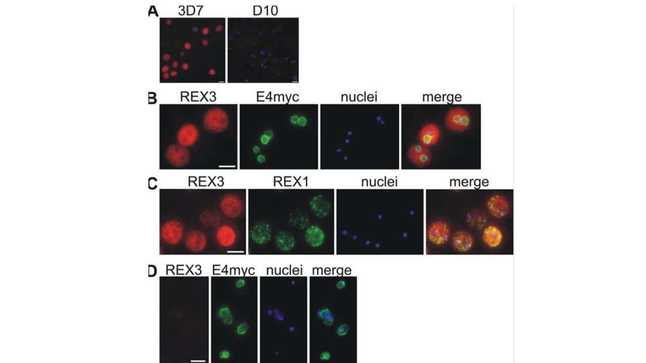
Location of REX3 in IRBCs fixed with 4% formaldehyde/0.005% glutaraldehyde. Acetone fixation was not suitable to detect REX3 (see Materials and Methods). (A) Specificity of the anti-REX3 serum: anti-REX3 serum reacted only with 3D7 (left) but not D10 parasites (right). Merges with nuclear stain (Hoechst) are shown. (B) IFAs with 3D7 parasites expressing a myc-tagged ETRAMP4 (Spielmann et al., 2006) showed that REX3 (red; Texas Red) occurs as a uniform stain outside of the PVM (ETRAMP4myc in the PVM detected by a rabbit anti-myc antibody; green, Cy2). The fainter red stain inside the area delineated by the PVM is likely to represent REX3 in the host cell cytoplasm from different focal planes above/below the parasite. No REX3 signal was detectable in D10 parasites expressing ETRAMP4myc (D). (C) REX3 (red; Texas Red) did not colocalize with the Maurer’s cleft protein REX1 (Hawthorne et al., 2004) (green; Cy2) in 3D7 parasites. Bars, 5 μm. A Cluster of Ring Stage–specific Genes Linked to a Locus Implicated in Cytoadherence in Plasmodium falciparum Codes for PEXEL-negative and PEXEL-positive Proteins Exported into the Host Cell. Spielmann T, Hawthorne PL, Dixon MW, Hannemann M, Klotz K, Kemp DJ, Klonis N, Tilley L, Trenholme KR, Gardiner DL. A cluster of ring stage-specific genes linked to a locus implicated in cytoadherence in Plasmodium falciparum codes for PEXEL-negative and PEXEL-positive proteins exported into the host cell. Mol Biol Cell. 2006 PMID: 16760427
= comp
Other associated proteins
| PFID | Formal Annotation |
|---|---|
| PF3D7_0936300 | ring-exported protein 3 |