Certain non-canonical PEXEL/HT variants are functional in a REX3 reporter.
The Plasmodium falciparum exportome contains non-canonical PEXEL/HT proteins
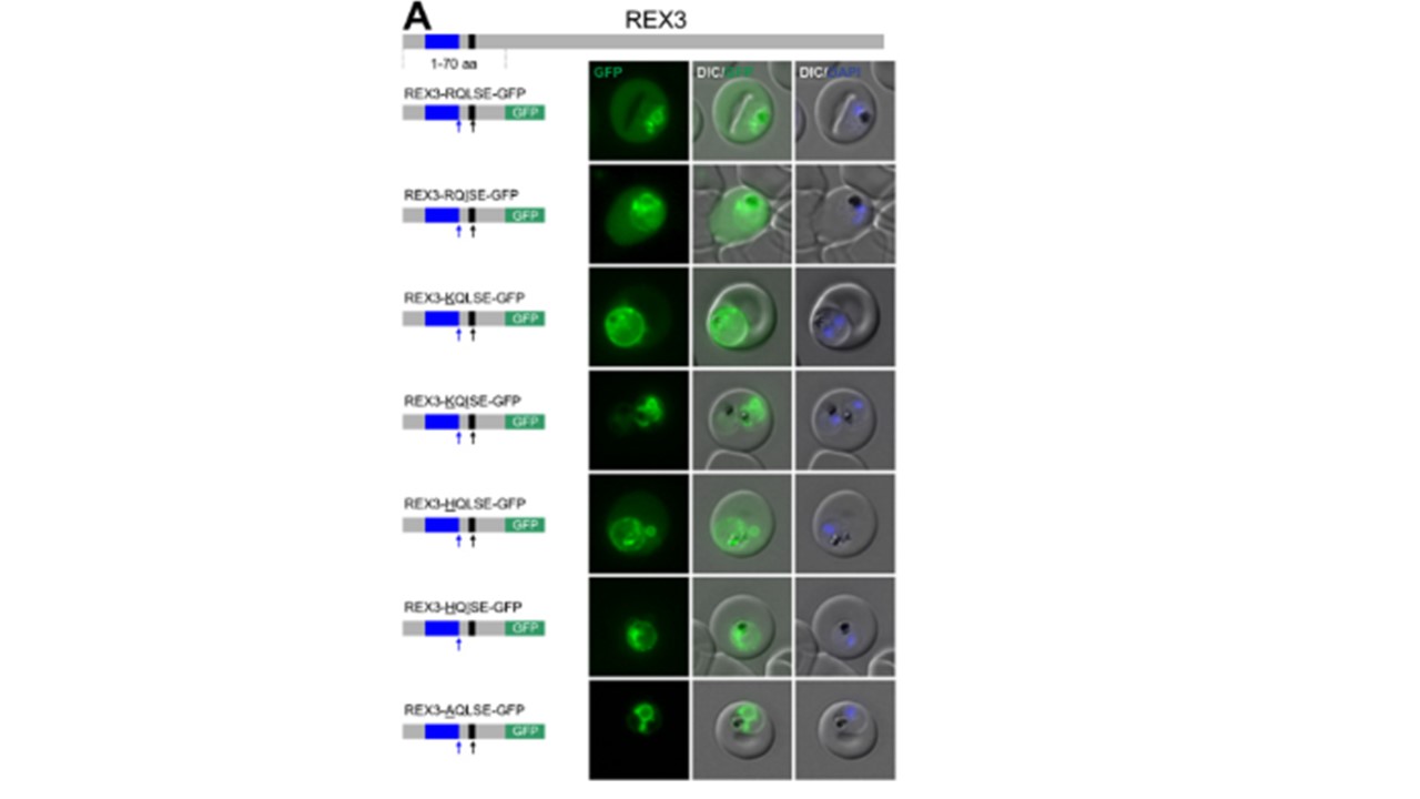
Fluorescence microscopy. The structure of full-length REX3 is indicated at the top; the schematic structure of the reporter proteins is depicted on the left of representative images of iRBCs. The ER-signal is shaded blue, the (non-) canonical PEXEL/HT is shaded black and GFP is shaded green. Putative cleavage sites are indicated by an arrow (blue: SP cleavage site; black: Plasmepsin V cleavage site). Fusion with the redox sensitive BPTI reveals a second translocation step for TM proteins. (A-D and F-L) Representative images of live P. falciparum parasites expressing the constructs shown schematically above each panel. Hydrophobic regions (SP, signal peptide; TM, transmembrane domain) are in black, the PEXEL motif in yellow. Numbers refer to amino acids (aa). Red boxes labelled C, additional REX2 C-termini. Interrupted yellow box, mutated BPTI (BPTImut). DIC, differential interference contrast. Size bars: 5 μm. (E) Schematic for the protease K (PK) protection assay. Left, intact infected RBC with 3 possibilities (I, II, III) for the location of the fusion construct: I, protein is integral to PVM; II, protein is freely accessible in the PV; III, protein is integral to PPM. Middle, after permeabilisation of the erythrocyte plasma membrane (EPM) with tetanolysin the N-terminus of the construct will be digested if it is in the PVM (I), but remains intact in situation II and III. Right, after permeabilisation of the PVM with saponin, the constructs will be digested if it is in the PVM (I) or the PV (II) but if in the PPM (III), an N-terminally truncated fragment will be generated. Red, exported protein; white box, TM; yellow, BPTI with double cysteine bonds; green, GFP. Schematic of full-length REX3 (top) and schematics and representative live microscopy images of reporter constructs containing the wild type recessed signal sequence or N-terminal truncation removing residues 2-17 to place the signal sequence immediately after the start methionine to mimic a classical signal peptide. SignalP 3 predicts a signal peptide for this arrangement with cleavage 14 residues upstream of the PEXEL. Scale bar: 5µm. Schulze J, Kwiatkowski M, Borner J, Schlüter H, Bruchhaus I, Burmester T, Spielmann T, Pick C. The Plasmodium falciparum exportome contains non-canonical PEXEL/HT proteins. Mol Microbiol. 2015 97(2):301-14. PMID: 25850860.