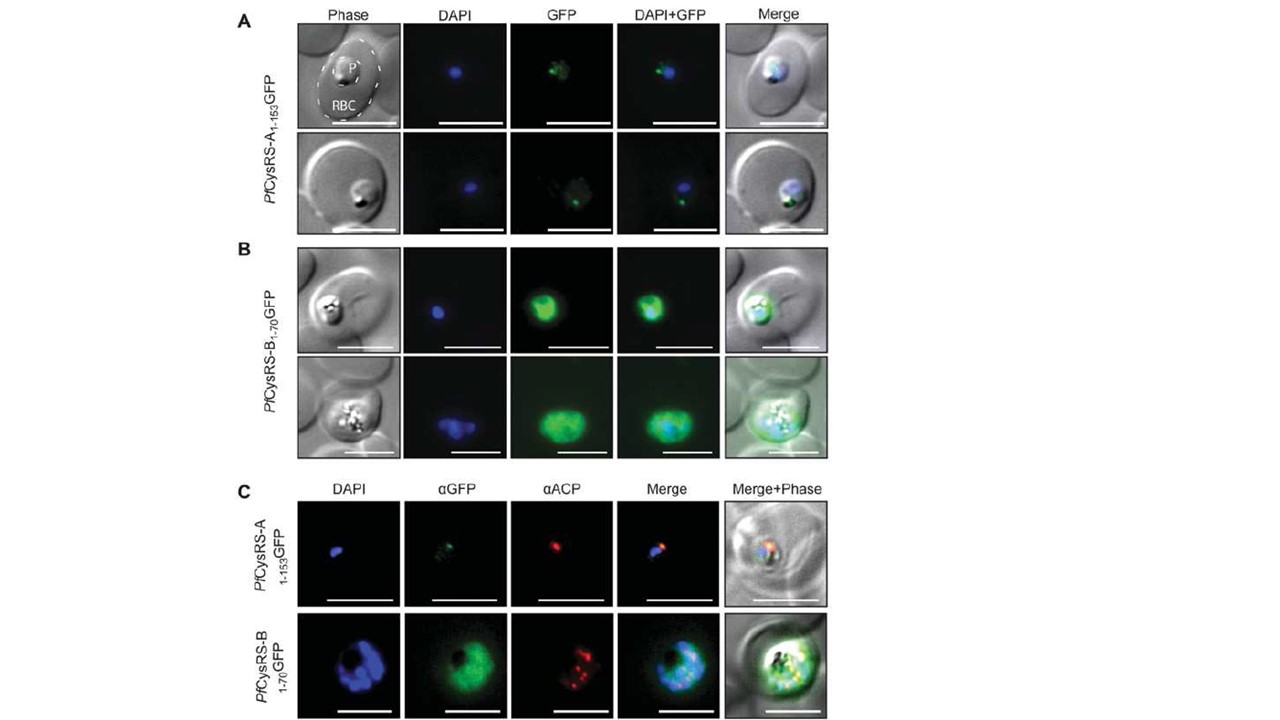
Localization of PfCysRS isoforms using GFP fusions in transgenic Plasmodium parasites Live-cell epifluorescence microscopy of P. falciparum transfected using pGlux.1 vector for episomal expression of GFP fused to the two PfCysRS mRNA isoforms: PfCysRS-A1–153GFP (A) and PfCysRS-B1–70GFP (B) in live cells. In (A), both parasites are early trophozoite stage, and localization of GFP is to a single punctum, consistent with the size and position of the apicoplast. The red blood cell (RBC) and the parasite (P) are indicated. (B) An early trophozoite (top panels) and late trophozoite (bottom panels) with GFP dispersed throughout the cytosol. (C) Immunofluorescence assays of the parasites indicated, PfCysRS-A1–153GFP (top panels) and Pf CysRS-B1–70GFP (bottom panels), using antibodies against GFP and ACP, an apicoplast marker. The PfCysRS-A1–153GFP signal specifically co-localizes with the apicoplast marker, whereas the Pf CysRS-B1–70GFP signal is distributed throughout the cytosol. For (A)–(C), all scale bars indicate 5 μm. (D) Western blot analysis of the transfectants indicated using an anti-GFP antibody. The two Pf CysRS–GFP fusions ran at their expected molecular mass, with PfCysRS-A1–153GFP in the mature processed form having the signal peptide and transit peptide cleaved off. The 3D7 parasite strain was used as a negative control and purified recombinant GFP was used as a positive control. Pham JS, Sakaguchi R, Yeoh LM, De Silva NS, McFadden GI, Hou YM, Ralph SA. A dual-targeted aminoacyl-tRNA synthetase in Plasmodium falciparum charges cytosolic and apicoplast tRNACys. Biochem J. 2014 458(3):513-23. PMID: 24428730
Other associated proteins
| PFID | Formal Annotation |
|---|---|
| PF3D7_0208500 | acyl carrier protein |