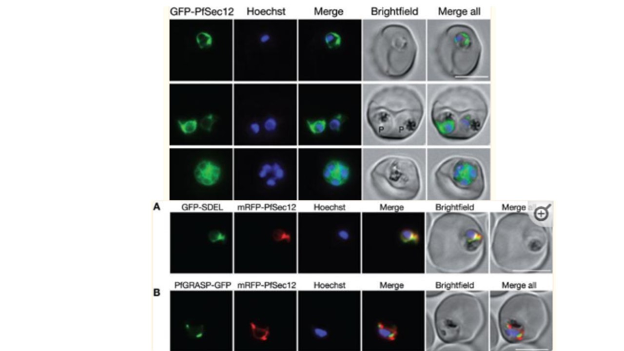
1. Live cell imaging of parasites expressing GFP-PfSec12 from the endogenous pfsec12 5′ UTR. Nuclei were stained with Hoechst 33342. GFP-PfSec12 is distributed in a perinuclear pattern with additional reticular protrusions characteristic of the parasite ER. Parasites at the ring, trophozoite and schizont stages are shown in the top, middle and bottom rows respectively. Note that the trophozoites (labelled P) were in a doubly infected erythrocyte. Bar = 5 μm.
2. Comparison of PfSec12 localization with markers for the ER and Golgi. Parasites were co-transfected with plasmids for expression of mRFP-PfSec12 (from the native pfsec12 5′ UTR) with either (A) GFP-SDEL, a marker for the general ER, or (B) PfGRASP-GFP, a cis-Golgi marker. mRFP-PfSec12 was localized throughout the ER coincident with GFP-SDEL, in close apposition to PfGRASP-labelled Golgi. Bar = 5 μm. Sun SY, Segev-Zarko L-a, Pintilie GD, Kim CY, Staggers SR, Schmid MF, Egan ES, Chiu W, Boothroyd JC. Cryogenic electron tomography reveals novel structures in the apical complex of Plasmodium falciparum. mBio. 2024 8:e0286423. PMID: 38456679.
Other associated proteins
| PFID | Formal Annotation |
|---|---|
| PF3D7_1116400 | guanine nucleotide-exchange factor SEC12 |