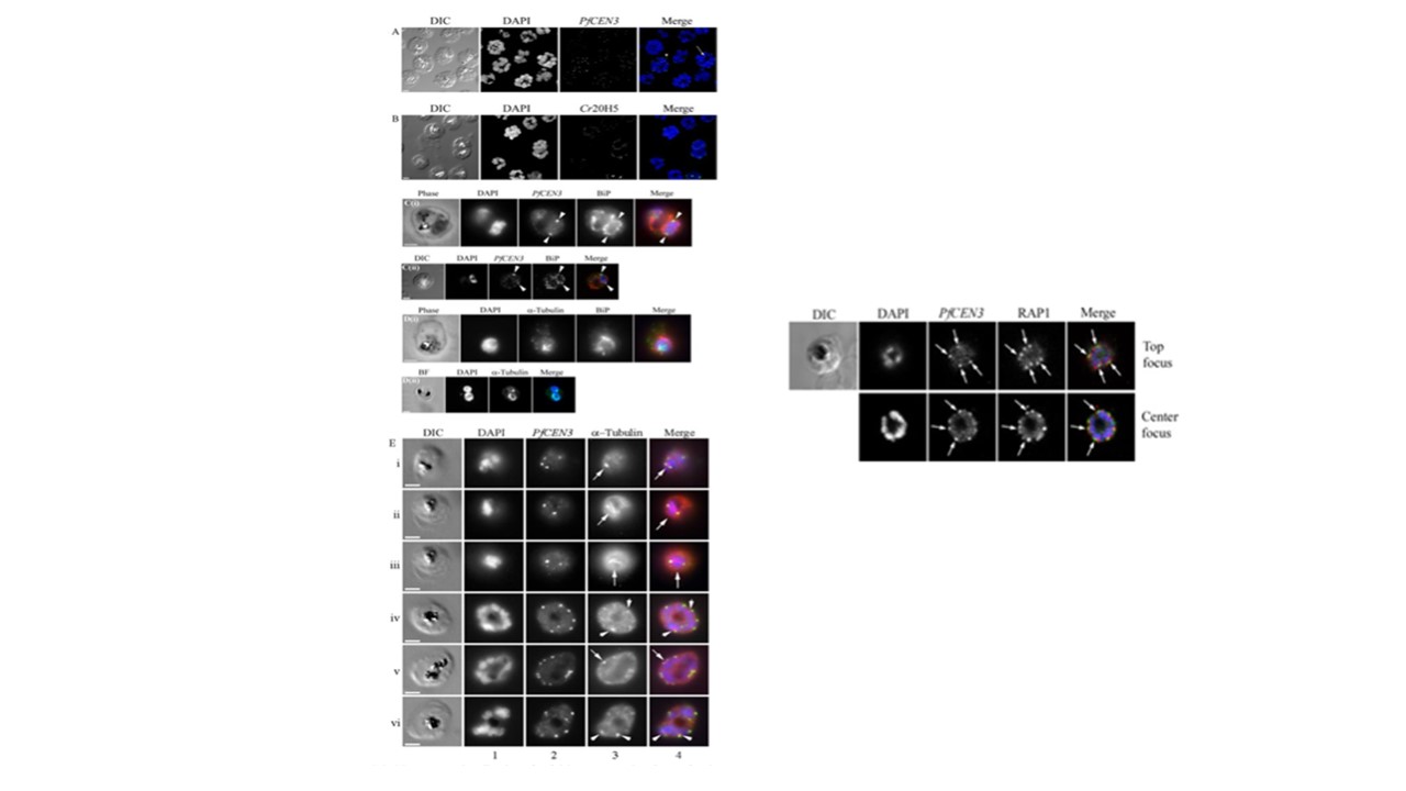
Left: Immunolocalization of P.falciparum centrin-3 by confocal and widefield epifluorescence microscopy. For confocal microscopy, P.falciparum asexual stage schizonts were stained with mouse anti-PfCEN3 antibody (A) and Cr20H5 monoclonal antibody (B). 1st panel depicts the differential interference contrast (DIC) image; 2nd panel depicts DAPI-stained nucleus (blue); the centrin reactivity is shown in the 3rd panel (green); and the 4th panel. Right:.Developing rhoptry pairs are arranged in a symmetrical pattern around PfCEN3 centrosomes. Confocal image of a schizont stage parasite. Nuclei are stained with DAPI (blue), anti-PfCEN3 antibody is labeled green, andanti-RAP1 antibody is labeled red to stainr hoptries. Rhoptries(red) are arranged in pairs around the peripheral cytoplasm of the developing schizont (RAP1, arrows). PfCEN3-positive centrosomes (green) are located between each rhoptry pair (Merge, arrows). Mahajan B, Selvapandiyan A, Gerald NJ, Majam V, Zheng H, Wickramarachchi T, Tiwari J, Fujioka H, Moch JK, Kumar N, Aravind L, Nakhasi HL, Kumar S. Centrins, cell cycle regulation proteins in human malaria parasite Plasmodium falciparum. J Biol Chem. 2008 283(46):31871-83.
