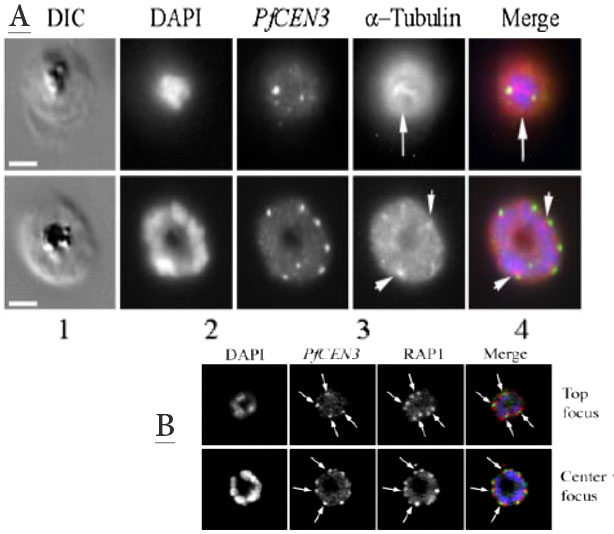
Upper panel: P. falciparum asexual stage schizonts were stained with a mixture of mouse anti-PfCEN3 (Alexa Fluor 488 label) and anti-a-tubulin PFI0180w (direct Alexa Fluor 680 label) antibodies or stained separately. DAPI staining the parasite nucleus is shown in blue channel in panel 1; PfCEN3 localization is shown on the green channel in panel 2; a-tubulin localization is shown on the red channel in panel 3. Merging of the blue, green, and red channels is shown on panel 4. The green dots represent the centrin localization to the poles of hemi-spindles and full spindles, the predicted localization of centrosomes (upper row). The association of centrin alongside of the DAPI-stained nucleus is shown in the lower row. Scale bar, 10 mm. When stained with PfCEN3 antibody alone, centrin reactivity appears as a pattern of paired spots that are organized around DAPI-positive nuclei.
Lower panel: Developing rhoptry pairs are arranged in a symmetrical pattern around PfCEN3 centrosomes. Confocal image of a schizont stage parasite. Nuclei are stained with DAPI (blue), anti-PfCEN3 antibody is labeled green, and anti-RAP1 antibody is labeled red to stain rhoptries. Rhoptries (red) are arranged in pairs around the peripheral cytoplasm of the developing schizont (RAP1, arrows). PfCEN3-positive centrosomes (green) are located between each rhoptry pair (Merge, arrows).
Mahajan B, Selvapandiyan A, Gerald NJ, Majam V, Zheng H, Wickramarachchi T, Tiwari J, Fujioka H, Moch JK, Kumar N, Aravind L, Nakhasi HL, Kumar S. Centrins, cell cycle regulation proteins in human malaria parasite Plasmodium falciparum. J Biol Chem. 2008 283:31871-83.
