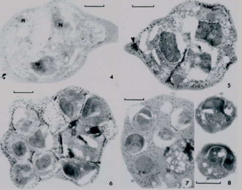
Electron micrographs from different parasite stages. 4. Early schizonts with 2 nuclei (N) and rhoprtry (R) and labeling over parasitophorous vacuole (PV). 5. Schizonts with labeled invaginating PV and with labeled vesicles within erythrocye cytoplasm (arrwohead). 6. Segmented schizont with labeled PV surrounding merozoites and residual body (containing pigment). Schizont with mature merozoites and PV membrane breakdown. Note even labeling of mixed RBC cytoplasm and PV contents. 8. Free merozoites in culture, free of specific label.
Culvenor JG, Crewther PE. S-antigen localization in the erythrocytic stages of Plasmodium falciparum. J Protozool. 1990 37:59-65. Copyright John Wiley & Sons Ltd. 2010.
