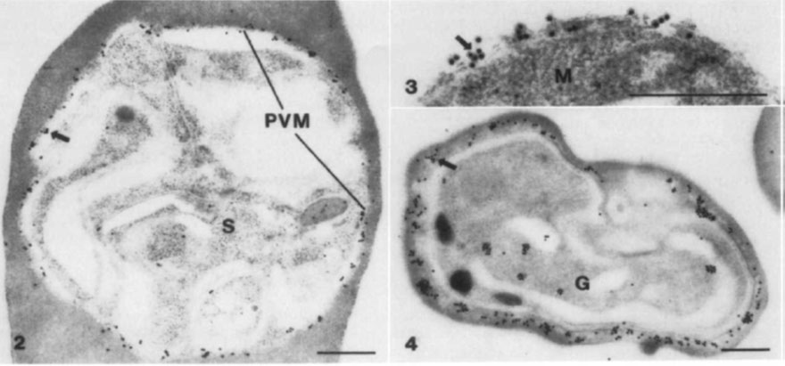
(2) Immuno-gold electron micrograph showing gold particles (arrow) distributed evenly along the parasitophorous vacuolar membrane (PVM) which surrounds a P. falciparum schizont (S). The presence of gold particles corresponds to the presence of GLURP. (3) immuno-gold electron micrograph of an erythrocytic merozoite (M). Staining (arrow) is on the surface of the merozoite. This indicates that GLURP is on the surface of the newly released merozoite, either as remnants of the parasitophorous vacuolar membrane or as a component of the merozoite membrane. Bar, 0.5 mm. (4) Gametocyte (G). lmmuno-gold staining (arrow) is located in clusters in the cytoplasm of the erythrocyte adjacent to the gametocyte.
Borre MB, Dziegiel M, Høgh B, Petersen E, Rieneck K, Riley E, Meis JF, Aikawa M, Nakamura K, Harada M, et al. Primary structure and localization of a conserved immunogenic Plasmodiumfalciparum glutamate rich protein (GLURP) expressed in both the preerythrocytic and erythrocytic stages of the vertebrate life cycle. Mol Biochem Parasitol. 1991 49:119-31. PMID: