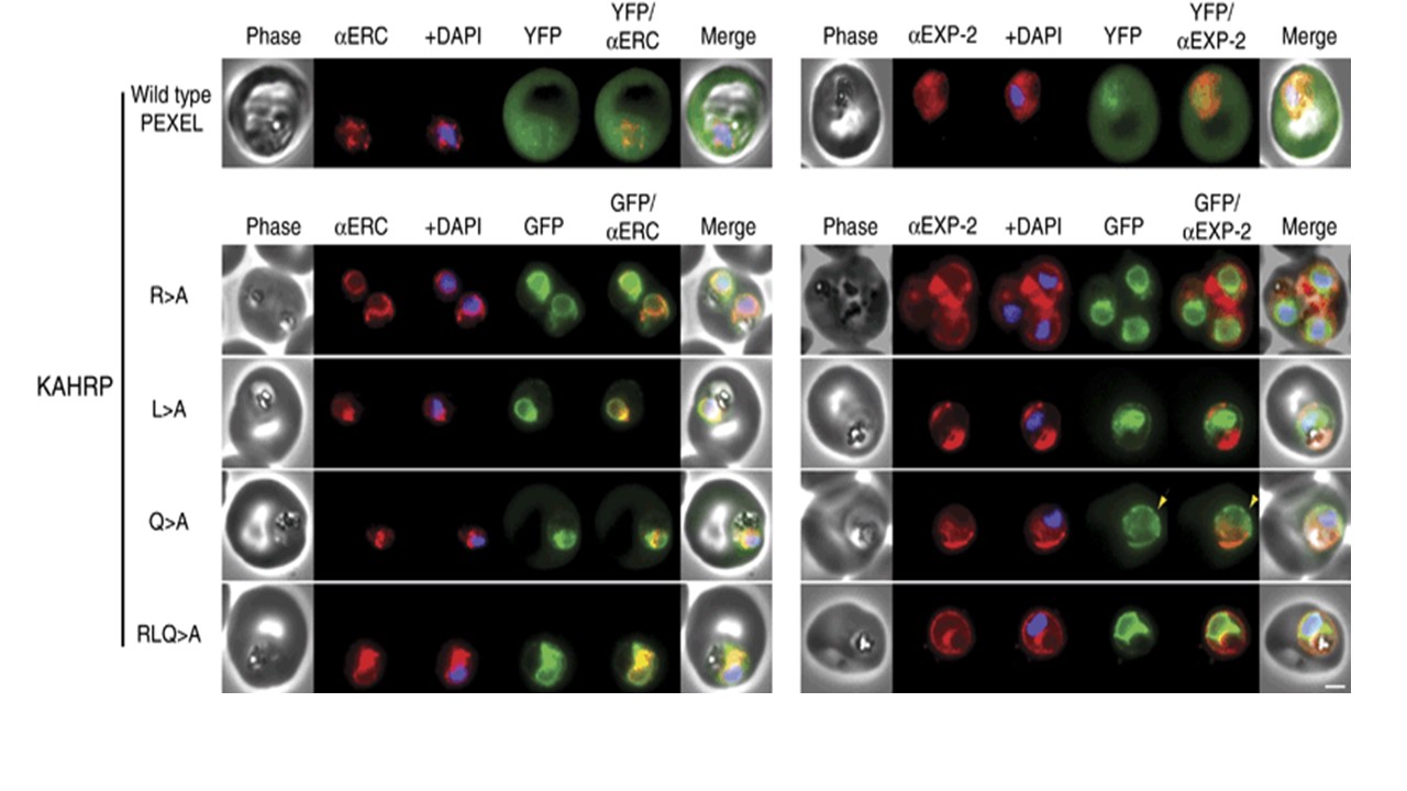
AHRP chimaeras localise to the endoplasmic reticulum in addition to the parasitophorous vacuole or erythrocyte cytosol. Images captured by immunofluorescence microscopy show substantial colocalisation of KAHRP chimaeras with the endoplasmic reticulum protein ERC (left panels). Intraparasitic fluorescence is also evident when chimaeras were colocalised with the parasitophorous vacuole membrane protein EXP-2 (right panels). Only the wild type (WT) PEXEL chimaera (uppermost panels) is efficiently exported to the erythrocyte cytosol, but low-level fluorescence in the erythrocyte cytosol was observed for the KAHRPQ>A chimaera. All mutants show some accumulation within the parasitophorous vacuole but while the KAHRPQ>A chimaera colocalises with ERC less than the other KAHRP mutants (left panel), it accumulates more in the parasitophorous vacuole in slightly older parasites (right panel). Accumulation of the KAHRPQ>A chimaera in the parasitophorous vacuole appeared inverse to the ‘necklace of beads’ observation described previously. The white bar in the last panel corresponds to 2 μm for each of the panels within the figure. Boddey JA, Moritz RL, Simpson RJ, Cowman AF. Role of the Plasmodium export element in trafficking parasite proteins to the infected erythrocyte. Traffic. 2009 10(3):285-99. PMID: 19055692.
Other associated proteins
| PFID | Formal Annotation |
|---|---|
| PF3D7_0202000 | knob-associated histidine-rich protein |
| PF3D7_1471100 | exported protein 2 |