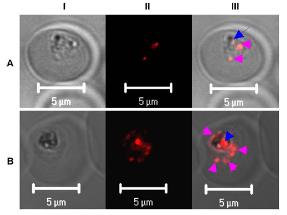
Immunolocalization of PfPRL in P. falciparum infected erythrocytes. The images represent the distribution patterns of PfPRL in P. falciparum intraerythrocytic stages. Synchronous 3D7 parasite cultures were probed with affinity purified rabbit anti-PfPRL followed by incubation with Alexa-Fluor 555-conjugated anti-rabbit IgG. (A) Two cytoplasmic foci (red arrowheads) and a single food vacuole-associated site (blue arrow) of anti-PfPRL reactivity are observed in mid-trophozoite stage parasites. (B) Schizont stage parasite showing increase in number of PfPRL-associted puncta during parasite development. Panel I is differential interference contrast image, panel II is fluorescence due to Alexa-Fluor 555 (red), and panel III is a merge of the first two panels.
Pendyala PR, Ayong L, Eatrides J, Schreiber M, Pham C, Chakrabarti R, Fidock DA, Allen CM, Chakrabarti D. Characterization of a PRL protein tyrosine
phosphatase from Plasmodium falciparum. Mol Biochem Parasitol. 2008 158(1):1-10. PMID: 18096253