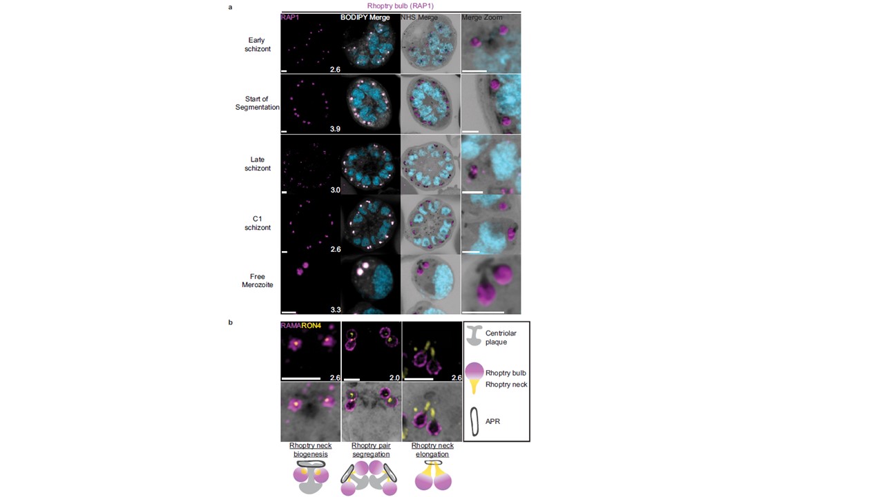
Rhoptries undergo biogenesis near the centriolar plaque and are segregated during nuclear division. 3D7 parasites were prepared by ultrastructural expansion microscopy (U-ExM), stained with N-hydroxysuccinimide (NHS) ester (grayscale), BODIPY TRc (white), SYTOX (cyan), and an anti-rhoptry antibodies and imaged using Airyscan microscopy. (a) Images of whole parasites throughout schizogony stained using an anti-RAP1 (rhoptry).
Liffner B, Cepeda Diaz AK, Blauwkamp J, Anaguano D, Frölich S, Muralidharan V, Wilson DW, Dvorin J, Absalon S. Atlas of Plasmodium falciparum intraerythrocytic development using expansion microscopy. 2023 PMID: 36993606;
PubMed Article: Atlas of Plasmodium falciparum intraerythrocytic development using expansion microscopy
Other associated proteins
| PFID | Formal Annotation |
|---|---|
| PF3D7_0105200 | RAP01 |