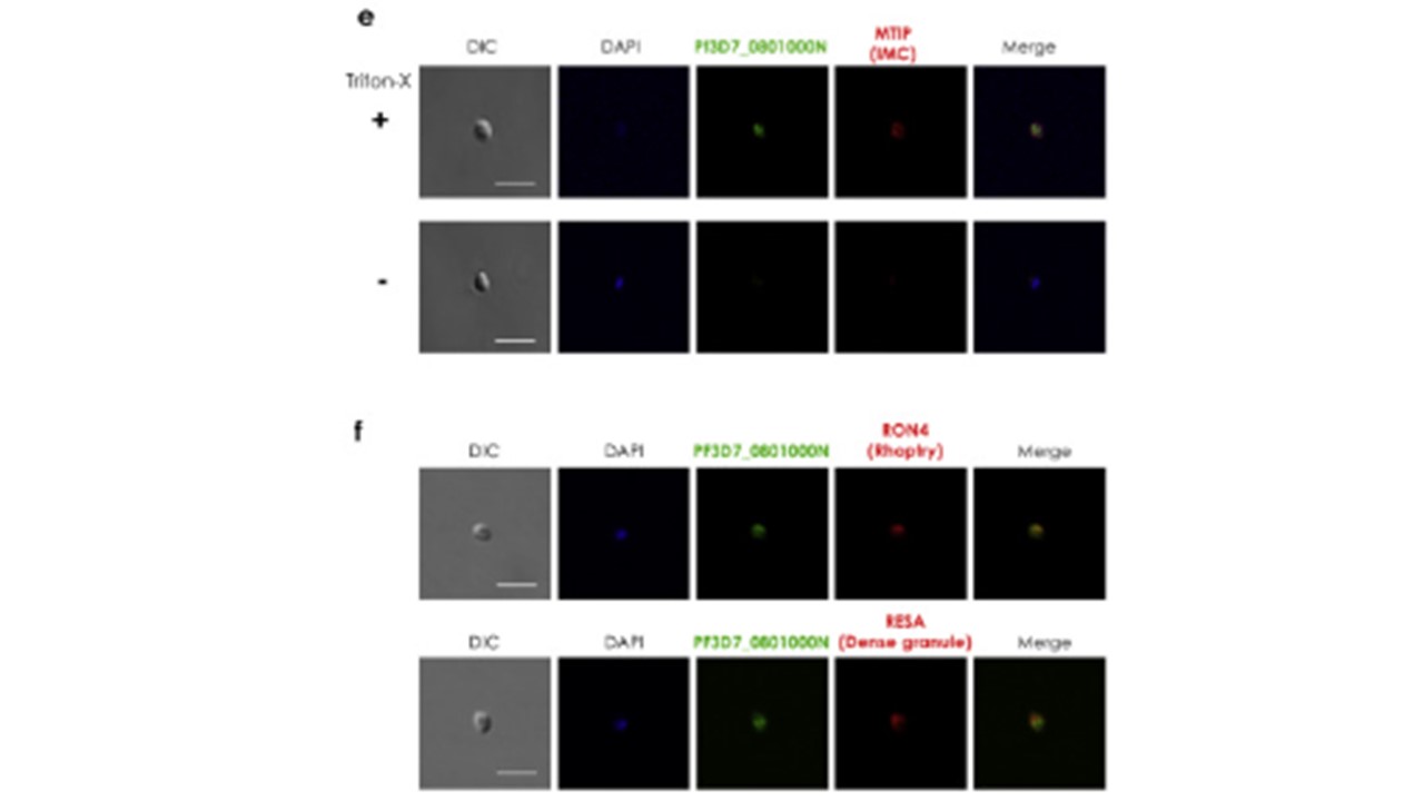
e. IFA image of PF3D7_0801000 relative to MTIP (myosin A tail domain interacting protein). Free merozoites were isolated as reported [13] and fixed at RT for 30 min with 4% paraformaldehyde/0.0075% glutaraldehyde in PBS. The merozoites were processed with (+) or without (−) 0.1% Triton X-100 permeabilization, followed by blocking with 3% bovine serum albumin (BSA) in PBS at RT for 30 min. Merozoites were co-stained with rabbit anti-PF3D7_0801000N antibodies and mouse anti-MTIP antibodies (marker for inner membrane complex (IMC) at 1:1000 and 1:100 dilutions, respectively. Alexa Fluor 488-conjugated goat anti-rabbit IgG and Alexa Fluor 568-conjugated goat anti-mouse IgG (Invitrogen, Carlsbad, CA) were used as secondary antibodies, at a 1:1000 dilution at RT for 30 min. DAPI (4′,6-diamidino-2-phenylindole) at 2 μg/ml was used to stain the nuclei. Merozoites were immobilized on polyethyleneimine-coated coverslips and mounted in ProLong Gold Antifade reagent (Invitrogen). The images were captured using confocal scanning laser microscope (LSM710, Carl Zeiss MicroImaging, Thornwood, NY) and processed by Image J (NIH). Other panels represent differential interference contrast (DIC), parasite nuclei localization (DAPI) and merged pictures (Merge). Scale bar = 3 μm.
f. IFA analysis of PF3D7_0801000 with RON4 and RESA. IFA analysis was conducted as described in the caption of Fig. 1e above. Free merozoites were stained with rabbit anti-PF3D7_0801000N antibodies (green); mouse monoclonal antibodies against RON4, a merozoite rhoptry marker (red, upper panel); or RESA, a marker for dense granules (red, lower panel). The antibodies against RON4 (PF3D7_1116000) and RESA (PF3D7_0102200) were kind gifts from Jean F. Dubremetz and Robin F. Anders, respectively. DAPI shows the localization of parasite nuclei while the leftmost and rightmost panels show DIC and merged images, respectively. Scale bar = 3 μm. Nagaoka H, Kanoi BN, Morita M, Nakata T, Palacpac NMQ, Egwang TG, Horii T, Tsuboi T, Takashima E. Characterization of a Plasmodium falciparum PHISTc protein, PF3D7_0801000, in blood- stage malaria parasites. Parasitol Int. 2021 PMID: 33147497.
r
Other associated proteins
| PFID | Formal Annotation |
|---|---|
| PF3D7_0102200 | ring-infected erythrocyte surface antigen |
| PF3D7_0801000 | Plasmodium exported protein PHISTc |