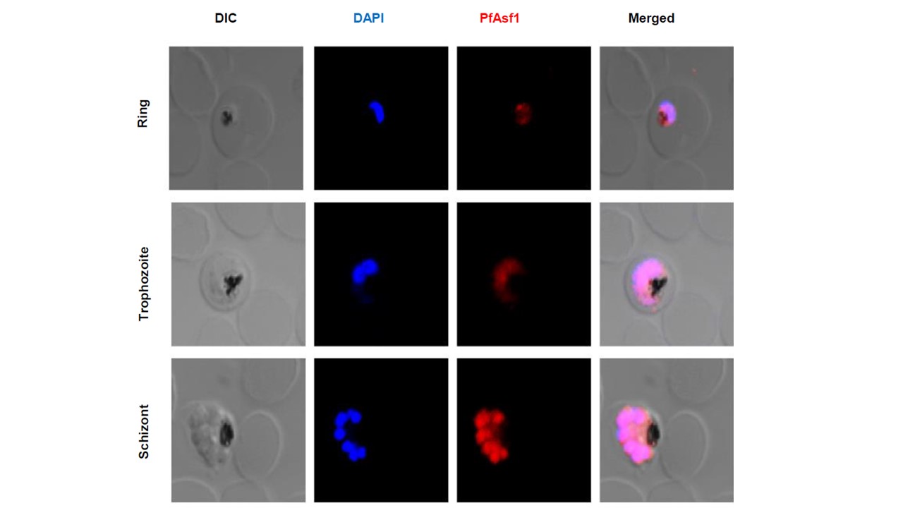
Stage-specific distribution of PfAsf1 in Plasmodium falciparum. Immunofluorescence based sub-cellular localization of PfAsf1 in different asexual blood stages (Ring, Trophozoite & Schizont) of P. falciparum. Shown in the panel are DIC (Differential Interference Contrast) images of the different stages of Plasmodium life cycle. DAPI (blue) was used as a nuclear marker and PfAsf1 was detected using anti-PfAsf1 antibody followed by secondary antibody conjugated to Alexa Flour 567(red). DIC, DAPI, PfAsf1 are merged in the fourth panel. These results indicate that PfAsf1 is ubiquitously present in different stages of Plasmodium life cycle and its expression reaches a peak at the Schizont stage.
Srivastava DK, Gunjan S, Das C, Seshadri V, Roy S. Structural insights into histone chaperone Asf1 and its characterization from Plasmodium falciparum. Biochem J. 2021 478(5):1117-1136.