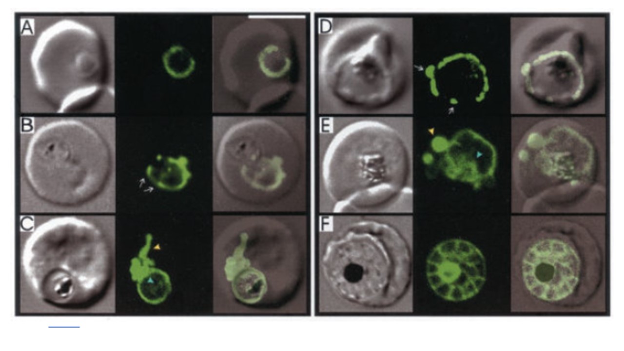
Expression of Exp1-(1–35)-GFP chimeric proteins at different stages of the intraerythrocytic life cycle of P. falciparum. The first image in each set represents the DIC image, and the second is the fluorescence signal from the GFP chimeric protein, with an overlay of these images in the third panel. A, ring stage parasite showing PV labeling. B, erythrocyte infected with two late ring stage parasites, only one of which is expressing the Exp1-(1–35)-GFP transgene. The fluorescence signal resembles a necklace of “beads,” some of which are marked with arrows. C–E, trophozoite stage parasites showing PV expression. Some cells retain the necklace of beads pattern (D). In some cases, distortions and evaginations of the PV were observed (C and E, yellow arrowheads). In some cells, GFP was observed in the FV (C and E, blue arrowheads). F, schizonts show a segmented pattern with a highly fluorescent central remnant body. Fluorescence from GFP was captured in live cells using a Leica TCS NT confocal microscope. Bar,5 mm. The Signal Sequence of Exported Protein-1 Directs the Green Fluorescent Protein to the Parasitophorous Vacuole of Transfected Malaria Parasites. Adisa A, Rug M, Klonis N, Foley M, Cowman AF, Tilley L. The signal sequence of exported protein-1 directs the green fluorescent protein to the parasitophorous vacuole of transfected malaria parasites. J Biol Chem. 2003 PMID: 12456681.
ºk