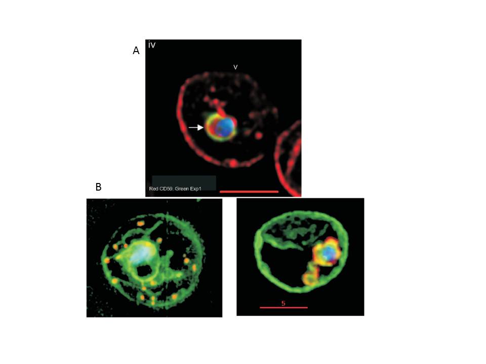
A. Internalization of host detergent-resistant membrane proteins in infected erythrocytes. Infected erythrocytes were probed with primary and relevant secondary antibodies to detect the indicated markers. CD59 (host cell marker), red; EXP1, green; . anti-CD59 antibody was directly conjugated to fluorescein isothiocyanate. The nucleus (blue) is stained with Hoechst, v indicates the periphery of the erythrocyte, arrows indicate vacuolar parasite, asterisks indicate tubular and vesicular membrane, and the scale bar is 5 mm.
B. Maurer’s clefts and intraerythrocytic loops are protein domains of the tubovesicular network. (Left) Infected cells labelled with BODIPY-ceramide (green) and probed with antibody Maurer’s cleft protein (red). Blue: parasite nucleus (Hoechst). (Right) Infected cells labelled with BODIPY-ceramide and probed with antibody to PfExp1 that detects intraerythrocytic loops (red). Blue: parasite nucleus (Hoechst).
Haldar K, Samuel BU, Mohandas N, Harrison T, Hiller NL. Transport mechanisms in Plasmodium-infected erythrocytes: lipid rafts and a tubovesicular network. Int J Parasitol. 2001 31:1393-401. Copyright Elsevier
