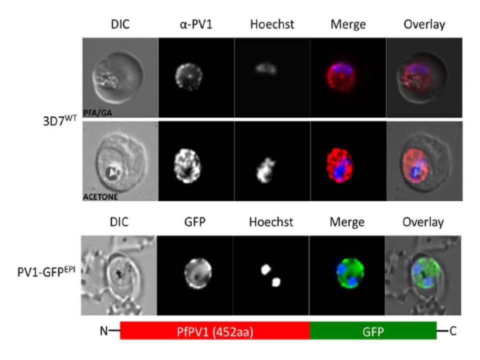
Localisation of PfPV1. (A, upper and middle panel) Immunofluorescence localisation using specific anti-PfPV1 antibodies. Lower panel: Episomal expression of a PfPV1-GFP chimera. DIC, differential interference contrast; GFP, green fluorescent protein; Hoechst, nuclear staining. GFP, green; specific antibody, red; Hoechst, blue. The imaging reveals GFP fluorescence in a ‘‘ring of beads’’ around the parasite, presumably the parasitophorous vacuole.
Chu T, Lingelbach K, Przyborski JM. Genetic evidence strongly support an essential role for PfPV1 in intra-erythrocytic growth of P. falciparum. PLoS One. 2011 6(3):e18396.