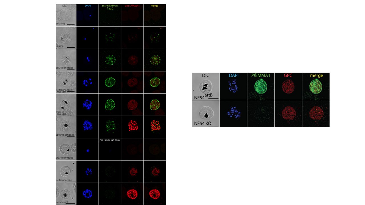
Left panel: Structural and temporal localization of PfEMMA1 by immunofluorescence confocal microscopy using anti-PfEMMA1 fragment 2 antisera. Representative permeabilized Pf3D7-infected RBCs were probed with mouse anti-PfEMMA1 fragment 2 (green) or preimmune sera and rabbit anti-PfAMA1 (red) and counterstained with DAPI to label parasite nuclei. PfEMMA1 is detected in all infected RBCs after the ring stage, when it is expressed in a stippled pattern outside the parasite, and then it aggregates at the RBC periphery but does not colocalize with PfAMA1, a merozoite marker. No proteins were detected with preimmune sera. Scale bar, 5 μm. DIC, differential interference contrast microscopy.
Right panel. Immunofluorescence confocal microscopy. Permeabilized RBCs infected with NF54attB or KO parasites at schizont stage were probed with mouse anti-PfEMMA1 fragment 2 and rabbit anti-GPC antibodies and counterstained with DAPI to label parasite nuclei.
Michelow IC, Park S, Tsai SW, Rayta B, Pasaje CFA, Nelson S, Early AM, Frosch AP, Ayodo G, Raj DK, Nixon CE, Nixon CP, Pond-Tor S, Friedman JF, Fried M, Duffy PE, Le Roch KG, Niles JC, Kurtis JD. A newly characterized malaria antigen on erythrocyte and merozoite surfaces induces parasite inhibitory antibodies. J Exp Med. 2021 218(9):e20200170.
Other associated proteins
| PFID | Formal Annotation |
|---|---|
| PF3D7_1134300 | erythrocyte membrane and merozoite antigen emma1 |