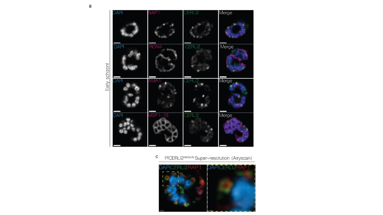
PfCERLI2 localises to the rhoptry bulb. a 2D Confocal immunofluorescence microscopy of early PfCERLI2HAGlmS schizonts stained with DAPI (nucleus) and anti-HA (PfCERLI2) antibodies, along with antibodies to either RAP1 (rhoptry bulb), RON4 (rhoptry neck), AMA1 (micronemes), or MSP1-19 (merozoite surface). Scale bar =2 μm.. c Representative maximum-intensity projection of PfCERLI2HAGlmS parasites stained with antibodies to RAP1 (rhoptry bulb) and HA (CERLI2) and imaged using the super-resolution microscopy platform Airyscan. Yellow box indicates the zoom area for the right-hand panel.
Liffner B, Balbin JM, Shami GJ, Siddiqui G, Strauss J, Frölich S, Heinemann GK, Edwards EM, Alder A, Wichers JS, Creek DJ, Tilley L, Dixon MWA, Gilberger TW, Wilson DW. Cell biological analysis reveals an essential role for Pfcerli2 in erythrocyte invasion by malaria parasites. Commun Biol. 2022 9;5(1):121. PMID: 35140336
Other associated proteins
| PFID | Formal Annotation |
|---|---|
| PF3D7_0405200 | cytosolically exposed rhoptry leaflet interacting protein 2 |
| PF3D7_0501600 | rhoptry-associated protein 2 |
| PF3D7_0930300 | merozoite surface protein 1 |
| PF3D7_1116000 | rhoptry neck protein 4 |