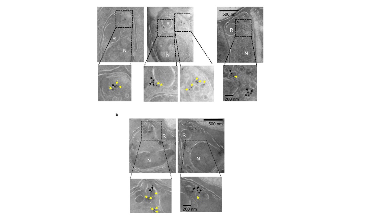
A subset of the apically located vesicles in schizonts showcolocalization
between AMA1 and PM X or SUB1 by immunoEM. Synchronized, C1-treated,
48–50 h schizonts expressing either PM X-3xHA (a) or SUB1-3xHA (b) were processed as described in the methods. Thin section samples were labeled for the indicated markers and visualized by immunoelectron microscopy. Black arrows: 18 nm beads that detect the anti HA antibody, yellow arrows: 12 nm beads that detect the anti AMA1 antibody. Sections showing apical vesicles aremagnified from each image. R rhoptry, N nucleus. Shown are representative images from two independent experiments.
Mukherjee S, Nguyen S, Sharma E, Goldberg DE. Maturation and substrate processing topography of the Plasmodium falciparum invasion/egress protease plasmepsin X. Nat Commun. 2022 Aug 4;13(1):4537. doi: 10.1038/s41467-022-32271-7. PMID: 35927261
Other associated proteins
| PFID | Formal Annotation |
|---|---|
| PF3D7_0507500 | subtilisin-like protease 1 |
| PF3D7_0808200 | plasmepsin X |