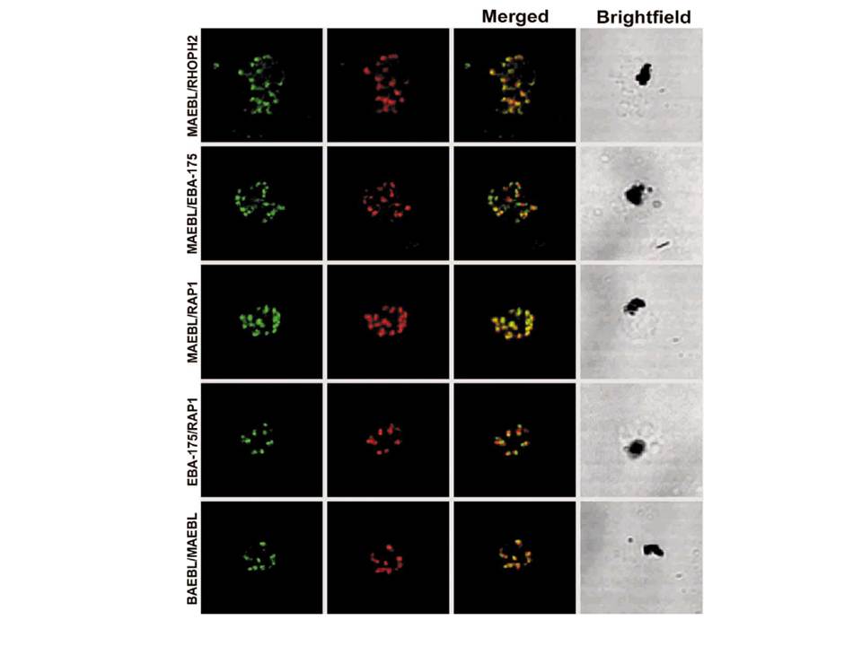
MAEBL localized with rhoptry proteins and on the merozoite surface and not with microneme proteins. Dual-label indirect immunofluorescence assays analyzed by scanning confocal laser microscopy are shown in each row of images, using primary antibodies against a rhoptry and microneme protein. In each row, the immunolocalization pattern for the first protein listed is in green in the first left-hand panel and the second protein listed is the second panel shown in red. The third panel is the merged image of the first two panels, the final right-hand panel is the brightfield image. Color was assigned electronically. Markers: RhopH2 PFI1445w; EBA-175 MAL7P1.176; RAP1 PFE0080c.
Blair PL, Kappe SH, Maciel JE, Balu B, Adams JH. Plasmodium falciparum MAEBL is a unique member of the ebl family. Mol Biochem Parasitol. 2002 122:35-44. Copyright Elsevier 2009.
Other associated proteins
| PFID | Formal Annotation |
|---|---|
| PF3D7_1301600 | erythrocyte binding antigen-140 |
