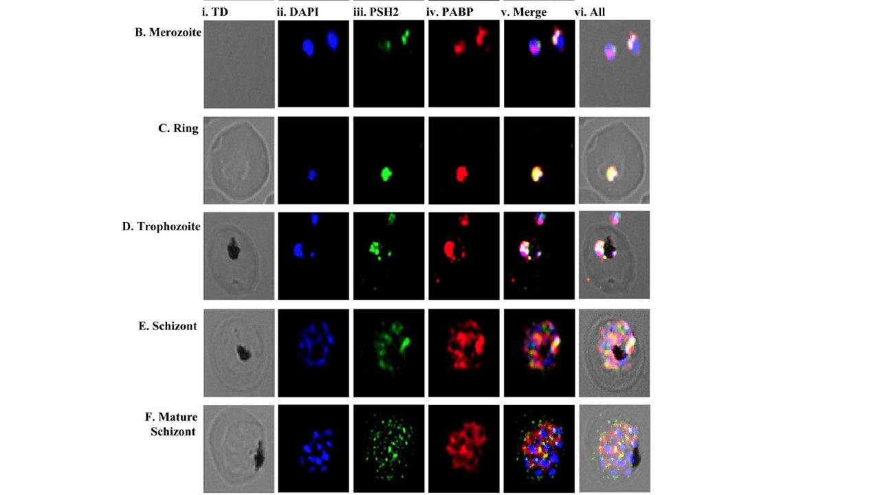
Localization of PSH2 in different intra-erythrocytic stages of P. falciparum. The cells were fixed and stained with pre-immune sera or anti-PfPSH2 antisera and anti-PfPABP antisera followed by Alexa fluor 488 and Alexa fluor 594-conjugated secondary antibodies and then counterstained with DAPI. In each panel, single confocal image of each stage is shown. (A) Staining with pre-immune sera (i) phase contrast (TD) image; (ii) image of cell stained with DAPI (blue); (iii) pre-immune sera PSH2; (iv) PSH2 + DAPI (v) All merged; (B–H) Staining with anti-PSH2 and anti-PABP sera (B) merozoite stage, (C) ring stage, (D) trophozoite stage, (E) schizont stage, (F) schizont stage (i) phase contrast (TD) image; (ii) image of cell stained with DAPI (blue); (iii) immunofluorescent stained cell (PSH2); (iv) immunofluorescent stained cell (PABP); (v) Merged image of panel ii, iii and iv; (vi) Merged image of panel i-v. The confocal images obtained using the anti-PfPSH2N sera suggest that the localization of PfPSH2 is mostly in the cytoplasm and it is very less in the nucleus at some intraerythrocytic developmental stages of the parasite, PfPSH2 and PfPABP colocalize in merozoite stages. Similarly, in ring stage the colocalization is even more visible. In case of trophozoite stage some nuclear localization of PfPSH2 is visible and in this stage of intraerythrocytic development, PfPSH2 also colocalizes with PfPABP. In schizont stages PfPSH2 shows punctate type localization that is scattered in the cytoplasm and it also colocalizes with PfPABP.
Chauhan M, Tuteja R. Plasmodium falciparum specific helicase 2 is a dual, bipolar helicase and is crucial for parasite growth. Sci Rep. 2019 9(1):1519.
Other associated proteins
| PFID | Formal Annotation |
|---|---|
| PF3D7_1224300 | polyadenylate-binding protein 1, putative |