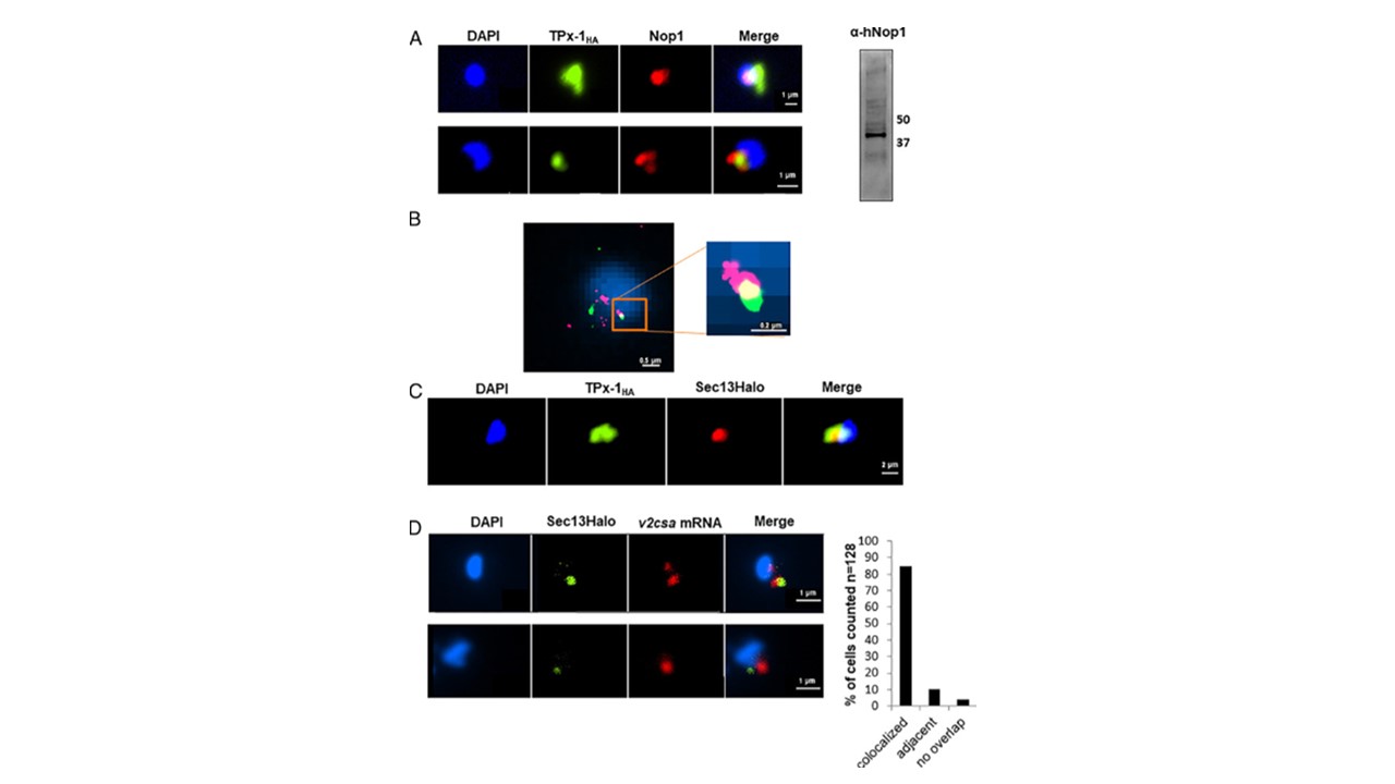
PfTPx-1 localizes to a transcriptionally active nuclear region in ring-stage parasites. (A, Left) Immunofluorescence microscopy of PfTPx-1HAglms parasites where PfTPx-1 (green) is associated with the nucleolar protein, PfNop1 (red). Nuclei are stained with DAPI. (Scale bar, 1 µm.) (Right) An anti hNop1/Fibrillarin antibody specifically detects PfNop1 in parasite extract. (B) Superresolution STORM imaging of PfTPx-1 (green) and PfNop1 (red) in synchronized ring-stage PfTPx-1HA parasites. Nuclei stained with YOYO1 were imaged by conventional epifluorescence for cellular orientation. An area of overlap is enlarged (square Inset). (Scale bar, 0.5 and 0.2 µm, respectively.) (C) Immunofluorescence of ring-stage PfTPx-1HAglms (PfTPx-1 in green) transfected with a Sec13-Halo plasmid (red). (Scale bar, 2 µm.) (D) RNA-FISH of CSA expressing parasites that carry a Sec13-Halo plasmid (green). RNA-FISH was performed using custom-designed Stellaris probes that hybridize to var2csa transcripts (red). Representative images of colocalized signals (Upper) and adjacent signals (Lower) are shown. (Scale bar, 1 µm.) Quantification of signals (n = 128) that were found to be overlapping or adjacent to each other are shown to the right. Heinberg A, Amit-Avraham I, Mitesser V, Simantov K, Goyal M, Nevo Y, Kandelis-Shalev S, Thompson E, Dzikowski R. A nuclear redox sensor modulates gene activation and var switching in Plasmodium falciparum. Proc Natl Acad Sci U S A. 2022 119(33):e2201247119.