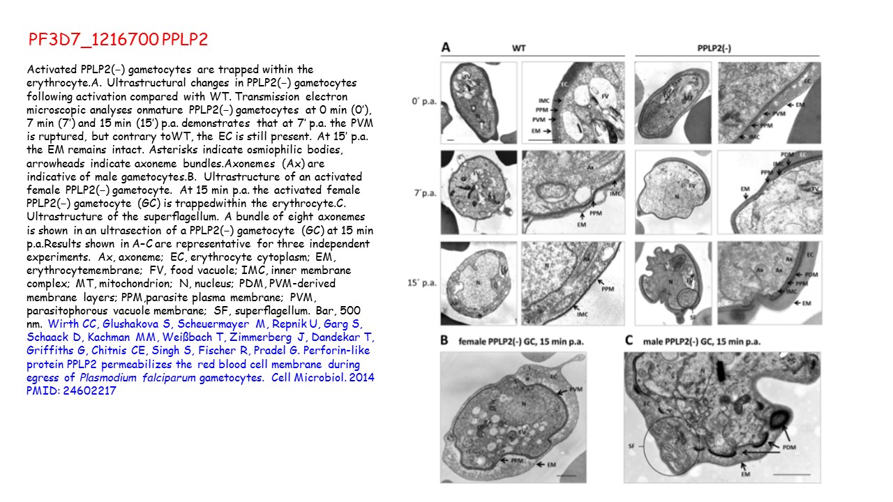
Activated PPLP2(−) gametocytes are trapped within the erythrocyte.A. Ultrastructural changes in PPLP2(−) gametocytes following activation compared with WT. Transmission electron microscopic analyses onmature PPLP2(−) gametocytes at 0 min (0′), 7 min (7′) and 15 min (15′) p.a. demonstrates that at 7′ p.a. the PVM is ruptured, but contrary toWT, the EC is still present. At 15′ p.a. the EM remains intact. Asterisks indicate osmiophilic bodies, arrowheads indicate axoneme bundles.Axonemes (Ax) are indicative of male gametocytes.B. Ultrastructure of an activated female PPLP2(−) gametocyte. At 15 min p.a. the activated female PPLP2(−) gametocyte (GC) is trappedwithin the erythrocyte.C. Ultrastructure of the superflagellum. A bundle of eight axonemes is shown in an ultrasection of a PPLP2(−) gametocyte (GC) at 15 min p.a.Results shown in A–C are representative for three independent experiments. Ax, axoneme; EC, erythrocyte cytoplasm; EM, erythrocytemembrane; FV, food vacuole; IMC, inner membrane complex; MT, mitochondrion; N, nucleus; PDM, PVM-derived membrane layers; PPM,parasite plasma membrane; PVM, parasitophorous vacuole membrane; SF, superflagellum. Bar, 500 nm. Wirth CC, Glushakova S, Scheuermayer M, Repnik U, Garg S, Schaack D, Kachman MM, Weißbach T, Zimmerberg J, Dandekar T, Griffiths G, Chitnis CE, Singh S, Fischer R, Pradel G. Perforin-like protein PPLP2 permeabilizes the red blood cell membrane during egress of Plasmodium falciparum gametocytes. Cell Microbiol. 2014 PMID: 24602217