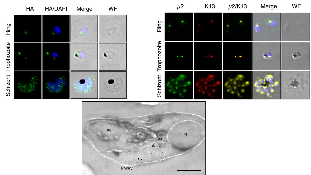
P. falciparum AP-2m is localized to a noncanonical cytoplasmic compartment. (Left panel) Localization of AP-2m-3xHA (green) across the asexual life cycle by anti-HA IFA, counterstained for parasite DNA with DAPI (blue). The images shown are representative of more than 100 cells examined at each stage; merge is the superimposition of each channel on a brightfield image (WF). Maximum intensity z-projections are shown. Scale bar, 2 mm. (Lower panel) Immunoelectron micrograph of a representative young intraerythrocytic trophozoite. AP-2m-3xHA parasites probed with an anti-HA rabbit antibody and a secondary antibody 18 nm gold conjugate. Protein disulfide isomerase (PDI), a marker for the parasite ER, is detected by an anti-PDI mouse antibody and a secondary conjugated to 12-nm gold particles. N, nucleus; FV, food vacuole; H, hemazoin; PM/PV, plasma membrane/parasitophorous vacuole; empty arrows, AP-2m associated with vesicles; black arrows, AP-2m at the plasma membrane; white-outlined arrows, AP-2m in the cytosol. Scale bar, 500 nm. (Right panel) Localization of AP-2m-3xHA (green) with respect to episomally expressed GFP-K13 (red) across the asexual life cycle by IFA. Representative images of more than 100 observed cells is shown. Maximum intensity z-projections are shown. Scale bar, 2 mm.
Henrici RC, Edwards RL, Zoltner M, van Schalkwyk DA, Hart MN, Mohring F, Moon RW, Nofal SD, Patel A, Flueck C, Baker DA, Odom John AR, Field MC, Sutherland CJ. The Plasmodium falciparum Artemisinin Susceptibility-Associated AP-2 Adaptin μ Subunit is Clathrin Independent and Essential for Schizont Maturation. mBio. 2020 Feb 25;11(1). pii: e02918-19.
