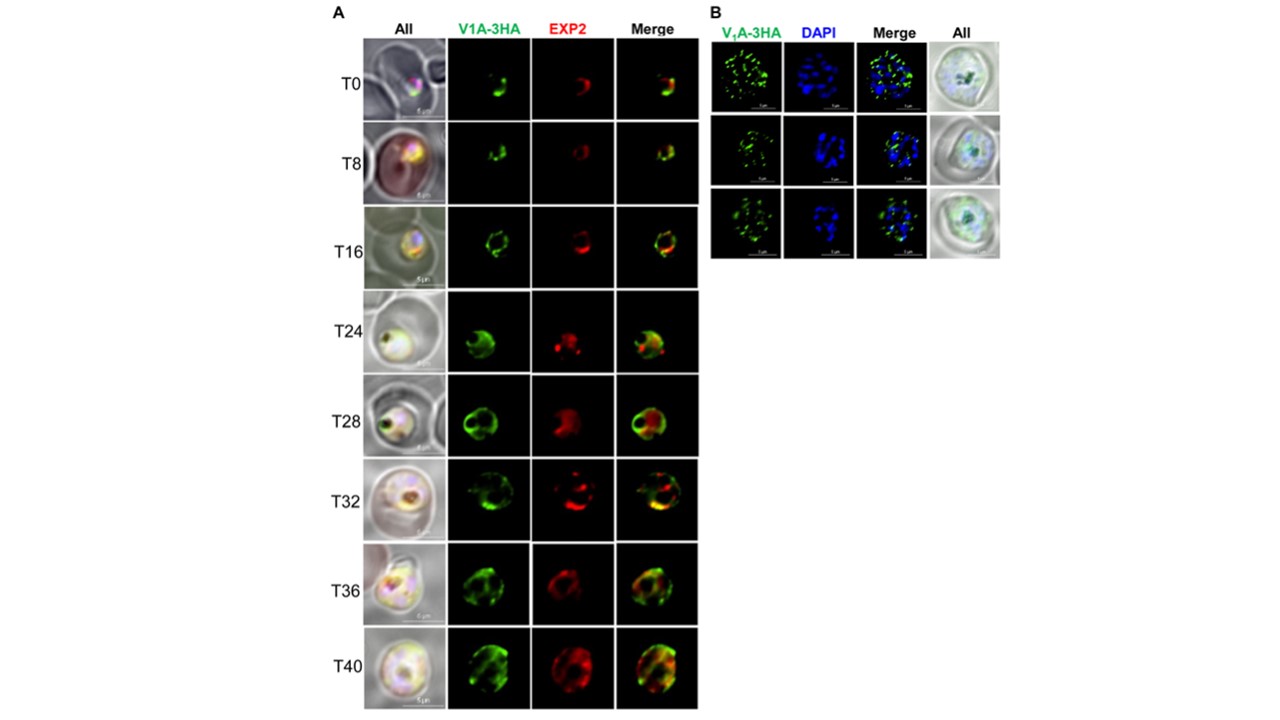
Localization of V1A throughout the asexual blood stages. A, immunofluorescence of the V1A protein in the ring, trophozoite, and schizont stages. In the NF54attB- V1A-3HAapt line, parasites were tightly synchronized and fixed every few hours starting from the early ring-stage (T0). V1A was detected using anti-HA and FITC-labeled secondary antibodies. Exp2, a marker for the parasitophorous vacuolar membrane (PVM), was detected using antiExp2 and tetramethylrhodamine-labeled secondary antibodies. B, immunofluorescence of V1A in mature schizont-stage parasites. The synchronized late trophozoite-stage parasites were treated with 25 nM of ML10 for 14 h to reach the mature schizont-stage (34). V1A was detected by anti HA and FITC-labeled secondary antibodies. In A-B, DNA was stained with 40,6-diamidino-2-phenylindole. 3HA, triple hemagglutinin epitope. Localization of V1A on secretory organelles in mature schizont-stage parasites.A–F, immuno-EM images of NF54attB- V1A -3HAapt in the mature schizont-stage. The synchronized late trophozoite-stage parasites were treated with 25 nM of ML10 for 14 h to reach the mature schizont-stage (34). Parasites were then fixed and subjected to immuno-EM analysis. V1A signals were detected on rhoptries (black arrows) and secretory organelles near rhoptries (red arrows). G and H, negative control images with the primary antibody omitted. Bars in A–H, 500 nm. 3HA, triple hemagglutinin epitope; Dv, digestive vacuole; Immuno-EM, immuno-electron microscopy; N, nucleus; PPM, parasite plasma membrane; PVM, parasitophorous vacuolar membrane; R, rhoptries; RBCM, red blood cell membrane; sv, secretory organelles. Shadija N, Dass S, Xu W, Wang L, Ke H. Functionality of the V-type ATPase during asexual growth and development of Plasmodium falciparum. J Biol Chem. 2024 300(9):107608. PMID: 39084459
Other associated proteins
| PFID | Formal Annotation |
|---|---|
| PF3D7_1471100 | exported protein 2 |
