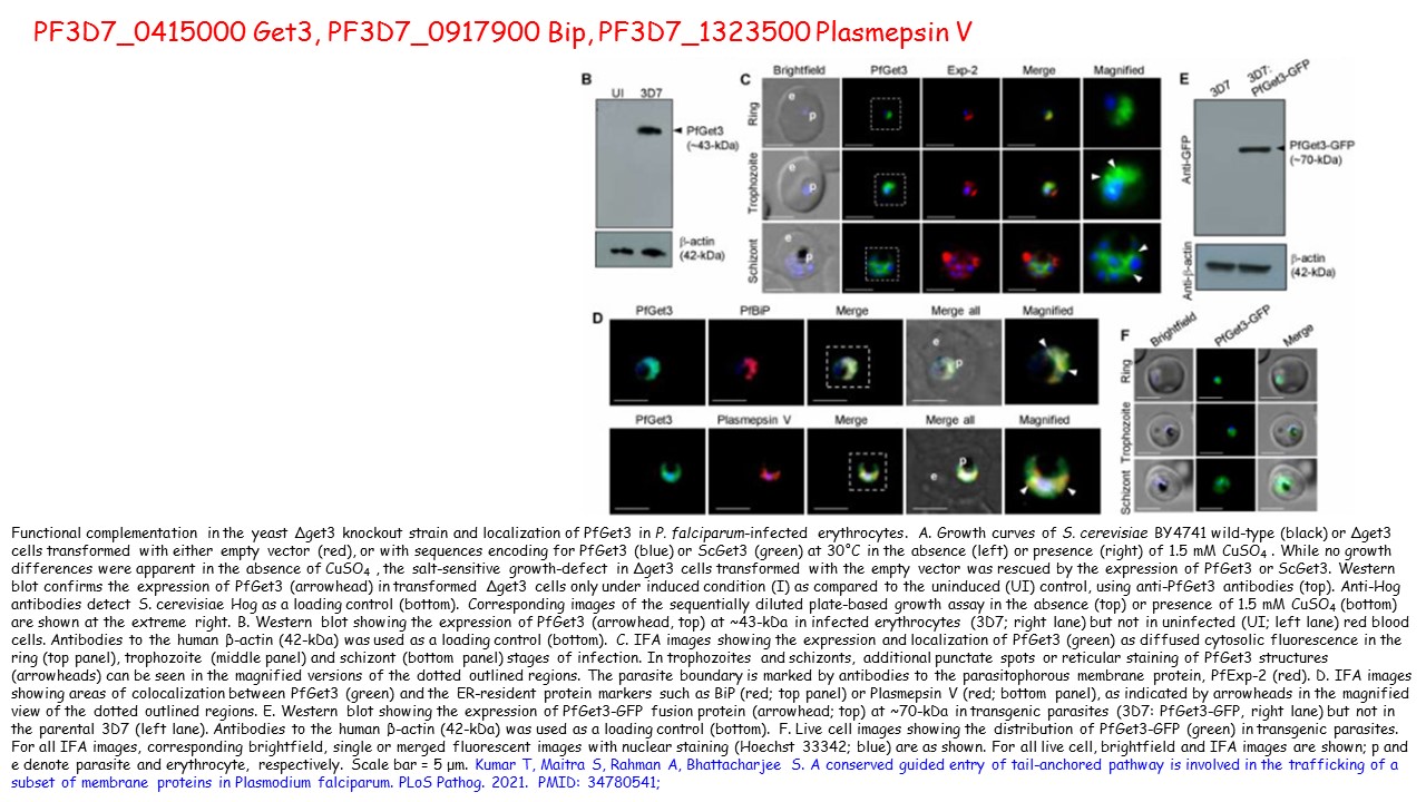
Functional complementation in the yeast Δget3 knockout strain and localization of PfGet3 in P. falciparum-infected erythrocytes. A. Growth curves of S. cerevisiae BY4741 wild-type (black) or Δget3 cells transformed with either empty vector (red), or with sequences encoding for PfGet3 (blue) or ScGet3 (green) at 30˚C in the absence (left) or presence (right) of 1.5 mM CuSO4 . While no growth differences were apparent in the absence of CuSO4 , the salt-sensitive growth-defect in Δget3 cells transformed with the empty vector was rescued by the expression of PfGet3 or ScGet3. Western blot confirms the expression of PfGet3 (arrowhead) in transformed Δget3 cells only under induced condition (I) as compared to the uninduced (UI) control, using anti-PfGet3 antibodies (top). Anti-Hog antibodies detect S. cerevisiae Hog as a loading control (bottom). Corresponding images of the sequentially diluted plate-based growth assay in the absence (top) or presence of 1.5 mM CuSO4 (bottom) are shown at the extreme right. B. Western blot showing the expression of PfGet3 (arrowhead, top) at ~43-kDa in infected erythrocytes (3D7; right lane) but not in uninfected (UI; left lane) red blood cells. Antibodies to the human β-actin (42-kDa) was used as a loading control (bottom). C. IFA images showing the expression and localization of PfGet3 (green) as diffused cytosolic fluorescence in the ring (top panel), trophozoite (middle panel) and schizont (bottom panel) stages of infection. In trophozoites and schizonts, additional punctate spots or reticular staining of PfGet3 structures (arrowheads) can be seen in the magnified versions of the dotted outlined regions. The parasite boundary is marked by antibodies to the parasitophorous membrane protein, PfExp-2 (red). D. IFA images showing areas of colocalization between PfGet3 (green) and the ER-resident protein markers such as BiP (red; top panel) or Plasmepsin V (red; bottom panel), as indicated by arrowheads in the magnified view of the dotted outlined regions. E. Western blot showing the expression of PfGet3-GFP fusion protein (arrowhead; top) at ~70-kDa in transgenic parasites (3D7: PfGet3-GFP, right lane) but not in the parental 3D7 (left lane). Antibodies to the human β-actin (42-kDa) was used as a loading control (bottom). F. Live cell images showing the distribution of PfGet3-GFP (green) in transgenic parasites. For all IFA images, corresponding brightfield, single or merged fluorescent images with nuclear staining (Hoechst 33342; blue) are as shown. For all live cell, brightfield and IFA images are shown; p and e denote parasite and erythrocyte, respectively. Scale bar = 5 μm. Kumar T, Maitra S, Rahman A, Bhattacharjee S. A conserved guided entry of tail-anchored pathway is involved in the trafficking of a subset of membrane proteins in Plasmodium falciparum. PLoS Pathog. 2021. PMID: 34780541;
ó
Other associated proteins
| PFID | Formal Annotation |
|---|---|
| PF3D7_0415000 | ATPase get3, putative, Get3 |
| PF3D7_0917900 | PfHsp70-2 |