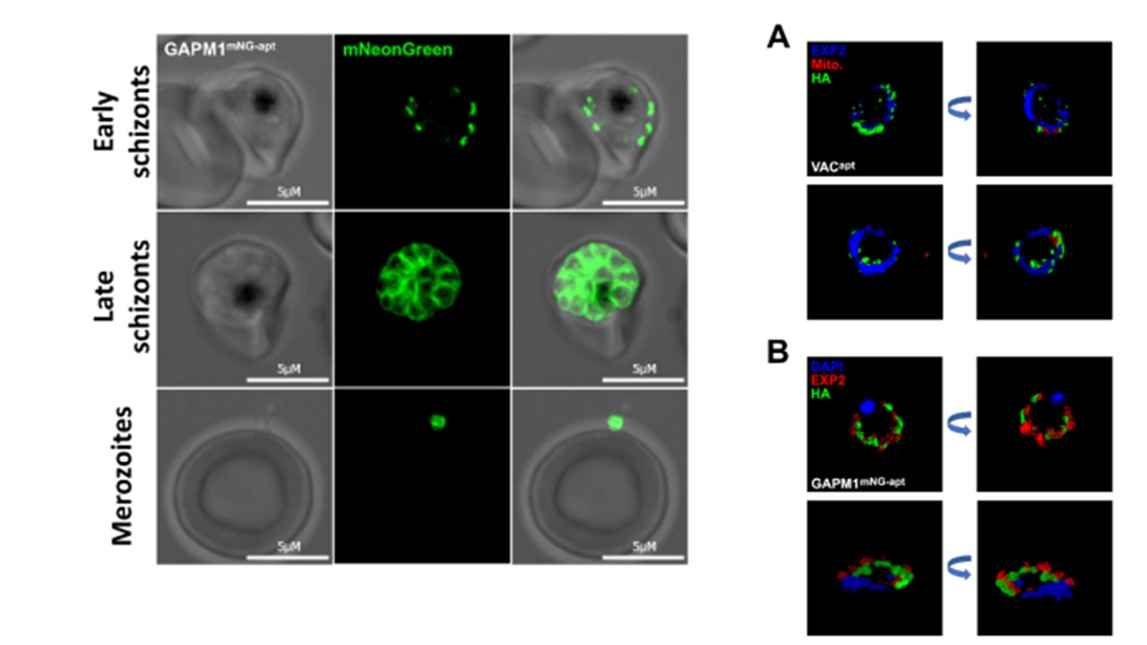
Left: Localization of GAPM1 at late stages in the GAPM1 mNG-apt cell line. (A) Representative live images showing GAPM1 mNG-apt localization at early and late schizonts, and merozoites. Images of GAPM1 mNG-apt from left to right are phase-contrast, mNeonGreen (green), and fluorescence merge. Z stack images were deconvolved and projected as a combined single image. Anaguano D, Dedkhad W, Brooks CF, Cobb DW, Muralidharan V. Time-resolved proximity biotinylation implicates a porin protein in export of transmembrane malaria parasite effectors. J Cell Sci. 2023 jcs.260506. PMID: 37772444.
PubMed Article: Time-resolved proximity biotinylation implicates a porin protein in export of transmembrane malaria parasite effectors
Other associated proteins
| PFID | Formal Annotation |
|---|---|
| PF3D7_1471100 | exported protein 2 |
