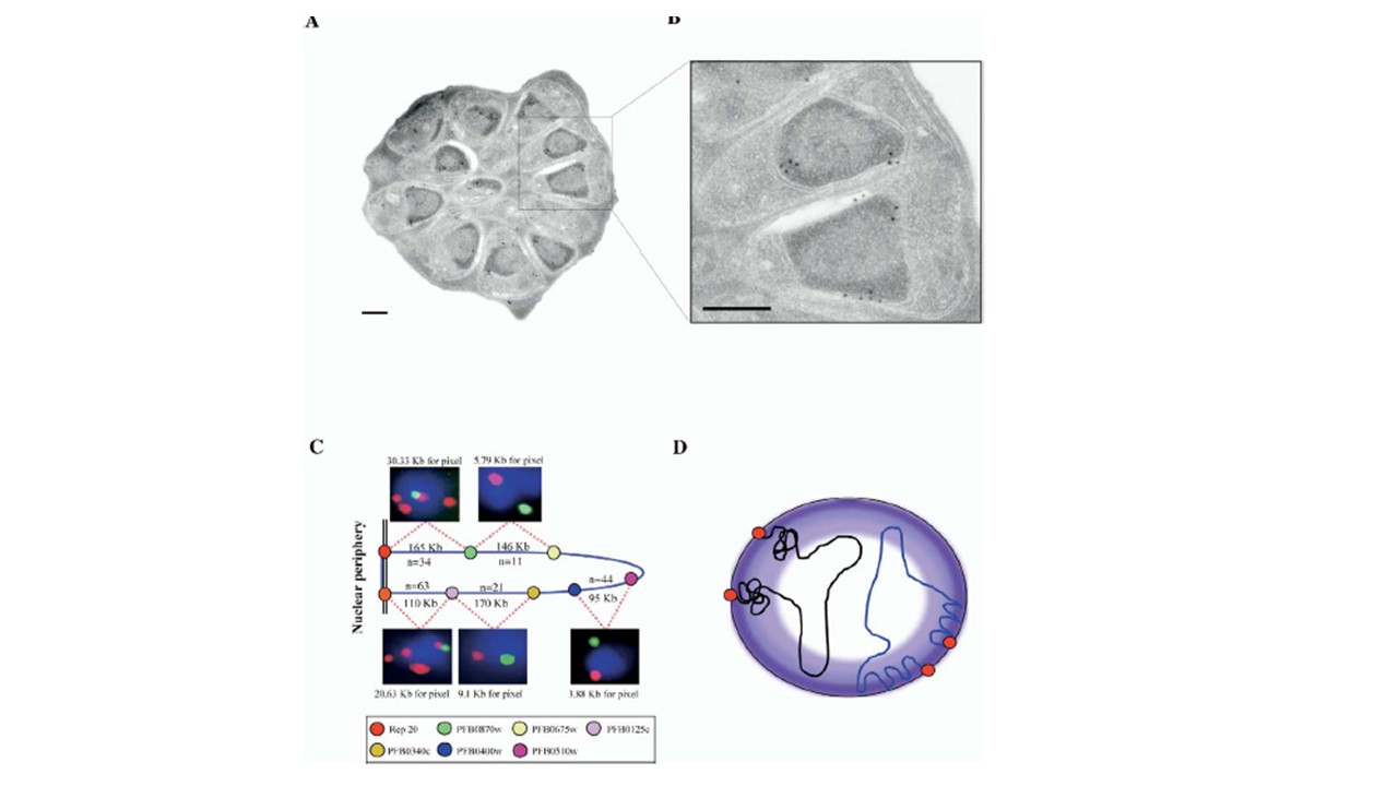
(A) The PfSir2 protein localizes to the electron-dense heterochromatic region at the nuclear periphery of P. falciparum parasites. (A) and (B) show developing merozoites in a late schizont-stage parasite. Scale bars in (A) and (B) are 250 mm.
(C) Schematic representation of FISH markers along P. falciparum chromosome 2. A closeup of the FISH signal is shown. The physical
distance between the markers is indicated as kilobases and values obtained using FISH analysis as kb/pixel. The probe names are indicated; n = number of observations.
(D) Model of organization of P. falciparum chromosome ends at the nuclear periphery, showing two possible chromatin conformations leading to compactness: heterochromatin or loop formation. Telomeres are shown as red dots.
Freitas-Junior LH, Hernandez-Rivas R, Ralph SA, Montiel-Condado D, Ruvalcaba-Salazar OK, Rojas-Meza AP, Mâncio-Silva L, Leal-Silvestre RJ, Gontijo AM, Shorte S, Scherf A. Telomeric heterochromatin propagation and histone acetylation control mutually exclusive expression of antigenic variation genes in malaria parasites. Cell. 2005. PMID: 15820676.
Other associated proteins
| PFID | Formal Annotation |
|---|---|
| PF3D7_1451400 | transcriptional regulatory protein sir2b |