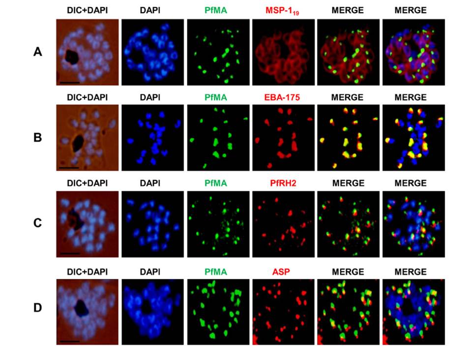
PfMA localization in schizont stages analyzed by fluorescence microscopy. Sub-cellular localization of PfMA was studied by co-immunostaining with surface protein (A), microneme (B), rhoptry (C, D) resident proteins. P. falciparum schizonts were co-immunostained with mouse anti-PfMA (green) and rabbit antibodies against one of the 4 marker proteins (EBA175/PfRH2, ASP, MSP-119) (red). The nuclei of schizont were stained with DAPI (blue) and slides were visualized by fluorescence microscope. All apical marker proteins and PfMA showed punctate staining in schizonts. PfMA was localized in the micronemes as it signal costained with micronemal marker EBA-175. The scale bar indicates 2 μm.
Hans N, Singh S, Pandey AK, Reddy KS, Gaur D, Chauhan VS. Identification and Characterization of a Novel Plasmodium falciparum Adhesin Involved in Erythrocyte Invasion. PLoS One. 2013 8(9):e74790.
Other associated proteins
| PFID | Formal Annotation |
|---|---|
| PF3D7_0316000 | microneme associated antigen |
| PF3D7_0405900 | apical sushi protein |
| PF3D7_0731500 | erythrocyte binding antigen-175 |
| PF3D7_0930300 | merozoite surface protein 1 |
| PF3D7_1335300 | reticulocyte binding protein 2 homologue b, ALKBH5 |
