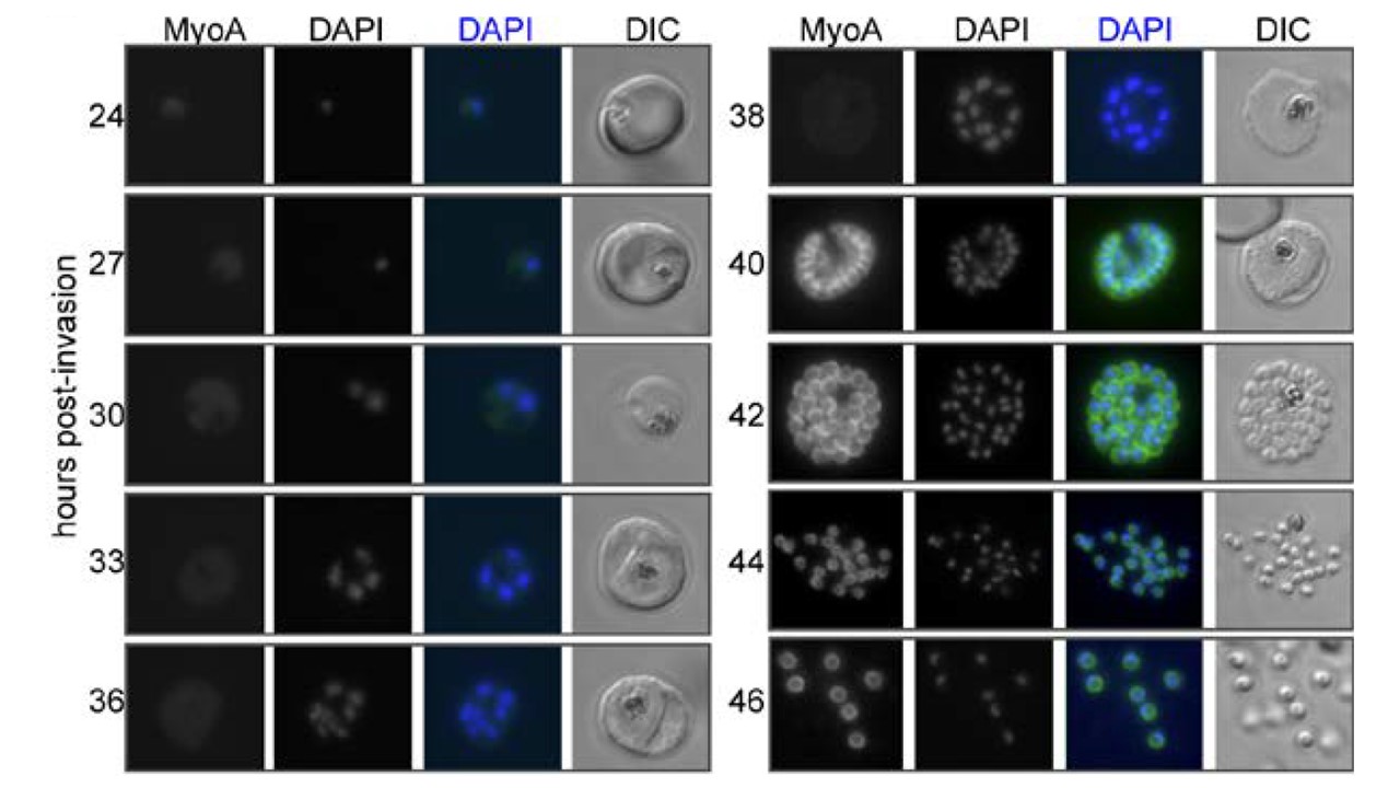
MyoA-GFP expression and glideosome complex formation during the late stages of intracellular development of P. falciparum in the red blood cell. MyoA-GFP expression was detected by live fluorescence microscopy and the nuclei were detected by staining with DAPI. The merged colour image with MyoA-GFP (green) and DAPI (blue) and differential interference contrast (DIC) bright field images are also shown. Schizogony starts at around 30 hours post-invasion and MyoA-GFP is detected from 38–40 hours in multinucleated forms. Scale bar: 2 μm.
Bookwalter CS, Tay CL, McCrorie R, Previs MJ, Lu H, Krementsova EB, Fagnant
PM, Baum J, Trybus KM. Reconstitution of the core of the malaria parasite
glideosome with recombinant Plasmodium class XIV myosin A and Plasmodium actin. J Biol Chem. 2017 Oct 4. .
