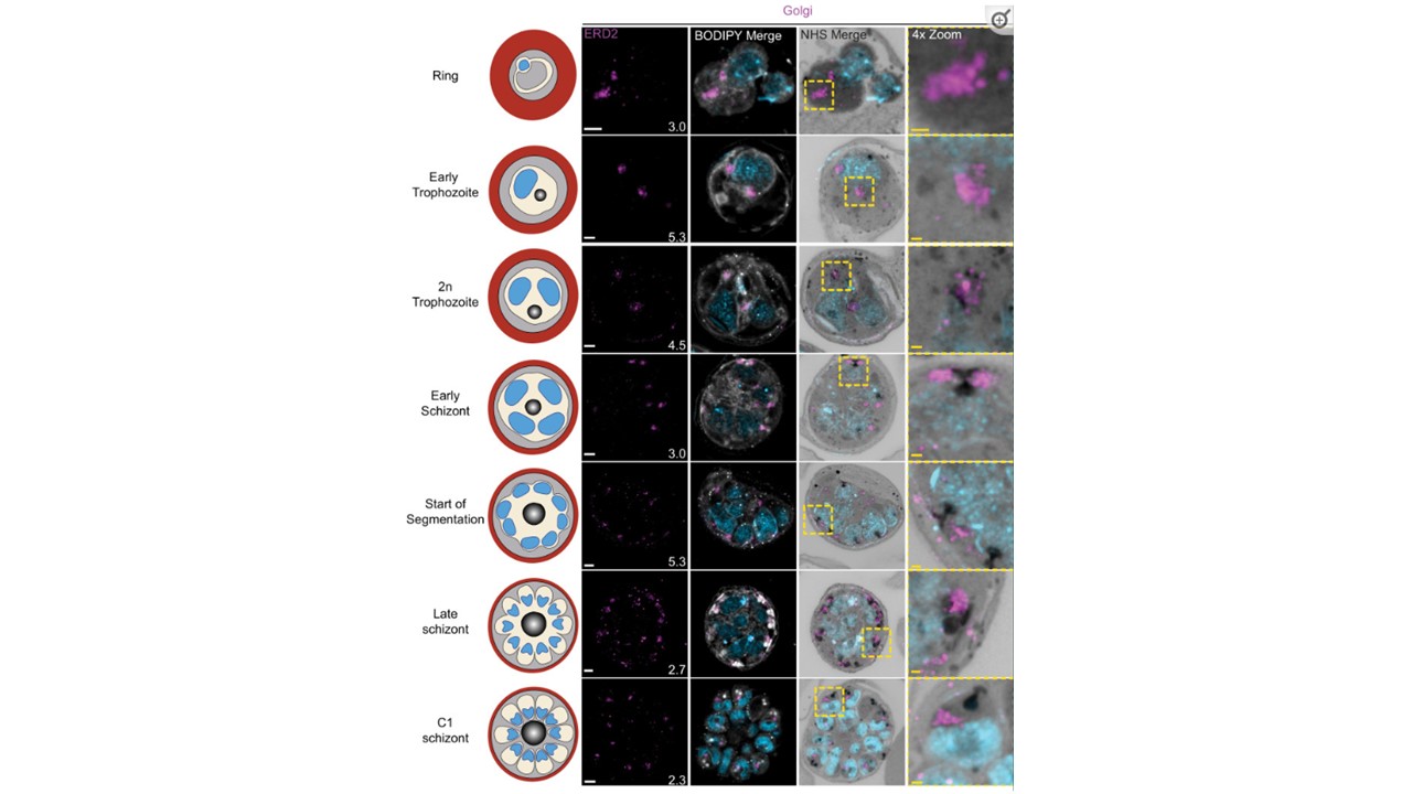
Golgi staining during intraerythrocytic development.
3D7 parasites were prepared by ultrastructural expansion microscopy (U-ExM), stained with N-hydroxysuccinimide (NHS) ester (grayscale), BODIPY TRc (white), SYTOX (cyan), and anti-ERD2 (Golgi; magenta) antibodies and imaged by Airyscan microscopy across the asexual blood stage. Images are maximum-intensity projections, number on image = Z-depth in µm of projection. White scale bars = 2 µm, yellow scale bars = 500 nm. 4× zooms show Golgi–centriolar plaque interaction. Liffner B, Cepeda Diaz AK, Blauwkamp J, Anaguano D, Frolich S, Muralidharan V, Wilson DW, Dvorin JD, Absalon S. Atlas of Plasmodium falciparum intraerythrocytic development using expansion microscopy. Elife. 2023 12:RP88088. PMID: 38108809