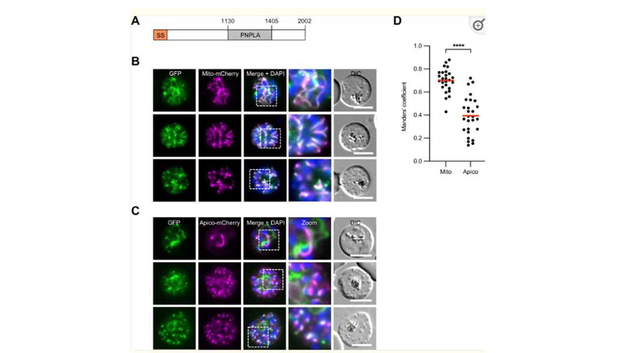
PfPNPLA2 localizes to the mitochondrion. (A) Schematic overview of the predicted functional domains of PfPNPLA2. SS, signal sequence; PNPLA, patatin-like phospholipase domain. Amino acid numbers are indicated. (B and C) Live-cell microscopy of parasites expressing endogenously tagged PfPNPLA2-GFP (green). Parasites co-expressing the mitochondrial marker Mito-mCherry (magenta) are shown in panel B, whereas parasites co-expressing the apicoplast marker Apico-mCherry (magenta) are shown in panel C. Merged images additionally contain DAPI-stained nuclei (blue). DIC, differential interference contrast. All scale bars, 5 µm. (C) Manders’ coefficients indicative of the fraction of PfPNPLA2-GFP overlapping the mitochondrial and apicoplast markers. Mean values from a total of 26 (Mito-mCherry) and 27 (Apico-mCherry) parasites are highlighted in red. For statistical analysis, unpaired Student’s t-test was used (****P < 0.0001). Pietsch E, Ramaprasad A, Bielfeld S, Wohlfarter Y, Maco B, Niedermüller K, Wilcke L, Kloehn J, Keller MA, Soldati-Favre D, Blackman MJ, Gilberger T-W, Burda P-C. A patatin-like phospholipase is important for mitochondrial function in malaria parasites. mBio. 2023 14(6):e0171823. PMID: 37882543;