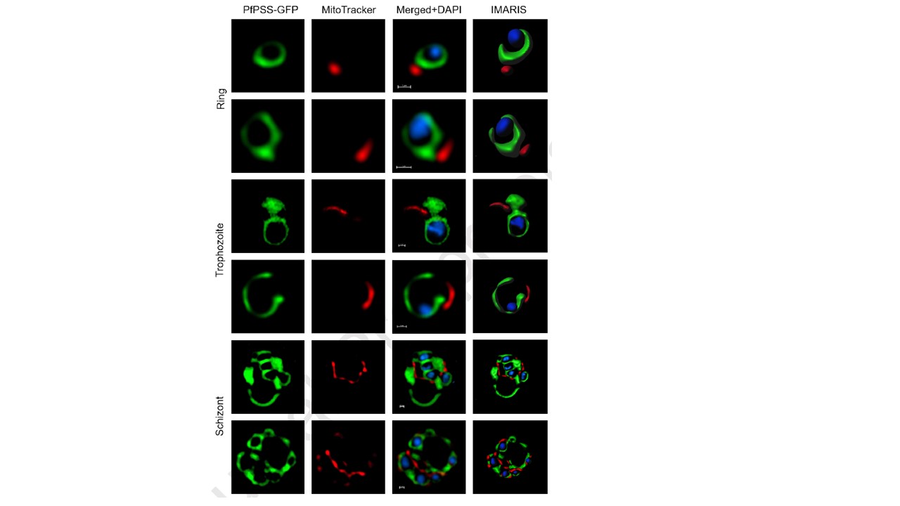
Representative Structured Illumination Microscopy (SIM) images of live PfPSS-GFP transgenic parasites showing association of ER and mitochondria in the asexual stages. For each SIM image, a three-dimensional reconstruction was developed using IMARIS software (shown in the right panel). Throughout the asexual stages, ER was found to be perinuclear with protrusions in cytoplasm, whereas mitochondrion in each of these parasites was closely associated with the ER. Mitochondrion is stained with MitoTracker Red and nuclei are stained with DAPI. In Merged+DAPI panel, scale bar = 1μm.
Anwar O, Islam M, Thakur V, Kaur I, Mohmmed A. Defining ER-mitochondria contact dynamics in Plasmodium falciparum by targeting component of phospholipid synthesis pathway, Phosphatidylserine synthase (PfPSS). Mitochondrion. 2022 :S1567-7249(22)00046-0.
