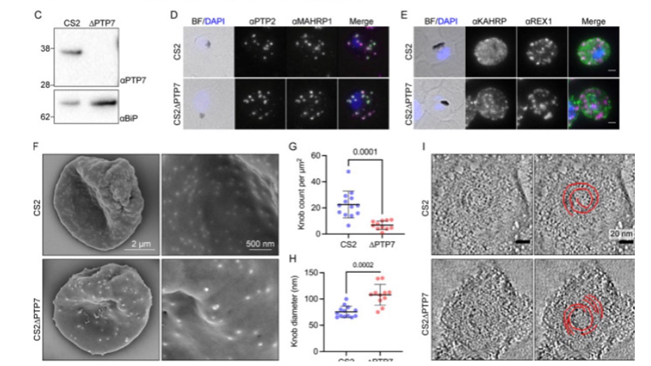
Disruption of the PTP7 locus affects knob and cleft morphology. (A) Schematic outlining the gene disruption strategy. DSB: double stranded break; RNP: ribonucleoprotein; yDHODH: yeast dihydroorotate dehydrogenase; Gray: coding sequence; up-caret: native intron; blue bulb and purple line: RNP and small guide RNA; HR1: homology region 1; HR2: homology region 2; crossing lines: homologous cross over events; arrows and letters A-D: primer locations. (B) PCR products of CS2 and CS2ΔPTP7 genomic DNA confirming disruption of the ptp7 locus. (C) Immunoblot of cell lysates probed with αPTP7. Loading control αBiP, expected size of ~62 kDa. (D-E) Indirect immunofluorescence microscopy of acetone/methanol fixed cells. Bright field (BF) and DAPI stained DNA (blue) images are merged. Scale bar, 2 μm. (F) Scanning electron microscopy of the exterior surface of mid-trophozoite stage infected RBCs. (G) Knob density as knob count per square μm, averaged per image. Data displayed are mean ± SD (n = 13, 10). (H) Data points represent the average knob diameter along the major axis for knobs in an individual cell. Data displayed are the mean ± SD (CS2 n = 13; ΔPTP7 n = 10). (I) Electron tomograms of infected RBCs membranes reveal the spiral structure underlying the knob. P-values determined by Welch’s t-test, n values are individual cells from ≥ 2 biological repeats. Carmo OMS, Shami GJ, Cox D, Liu B, Blanch AJ, Tiash S, Tilley L, Dixon MWA. Deletion of the Plasmodium falciparum exported protein PTP7 leads to Maurer's clefts vesiculation, host cell remodeling defects, and loss of surface presentation of EMP1. PLoS Pathog. 2022 18(8):e1009882. PMID: 35930605
Other associated proteins
| PFID | Formal Annotation |
|---|---|
| PF3D7_0202000 | knob-associated histidine-rich protein |
| PF3D7_0301700 | Plasmodium exported protein, unknown function |
| PF3D7_0935900 | ring-exported protein 1 |
