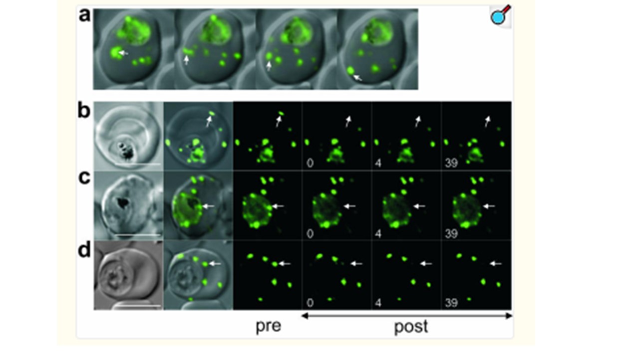
Molecular organization of GFP chimeras in different compartments. (a) Overlay images of a MAHRP11-130-GFP transfectant displaying mobile Maurer's clefts taken at consecutive time points. One moving cleft observed over time is indicated with white arrows. A movie of this cell and other examples can be viewed in Fig. S4. The bar represents 5 μm. (b to d) Photobleach analysis. The panels show the DIC images and the prebleach (pre) and postbleach (post) fluorescence images at the times (in seconds) indicated. The position of the bleach pulse is indicated by white arrows. (b) Bleaching of MAHRP152-249-GFP in a tethered peripheral Maurer's cleft with a 300-ms bleach pulse. There is no recovery of the signal after photobleaching, indicating a physically separate structure. Bleaching of a PV membrane-associated MAHRP152-249-GFP focal region with 300-ms bleach pulses (c) and a structure in close proximity to the PV membrane with a 200-ms bleach pulse (d). There is no recovery of fluorescence into the bleached areas. Scale bars, 5 μm. Spycher C, Rug M, Klonis N, Ferguson DJ, Cowman AF, Beck HP, Tilley L. Genesis of and trafficking to the Maurer's clefts of Plasmodium falciparum-infected erythrocytes. Mol Cell Biol. 2006. PMID: 16705161