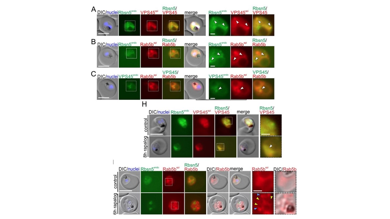
PfRab5b and PfVPS45 interact with PfRbsn5. A-C, Live-cell microscopy images of trophozoites of PfRbsn5endo parasites co-expressing PfVPS45epi (A), PfRab5bepi (B) or VPS45endo parasites, co-expressing PfRab5bepi (C). White arrows: overlapping of PfRbns5endo or PfVPS45endo and PfRab5bepi signals.
H, Live-cell microscopy images of PfRbsn5 knock-sideway (8 h rapalog) and
controls using the PfRbsn5-2x2/NLSendo parasites co-expressing PfVPS45epi. Representative images of n = 10 (control) and n = 15 (rapalog) cells. White arrow: Localization of PfVPS45 and Rbsn5 to the nucleus upon PfRbsn5 inactivation. I, Live-cell microscopy images of Rbsn5 knock-sideway (6 h rapalog) and controls of PfRbsn5-2x2/NLSendo parasites co-expressing PfRab5bepi . Yellow arrows: PfRab5bepi signal surrounding vesicles observed by DIC. Blue arrows: PfRab5b accumulations. Representative images of n = 2 independent experiments with 11, 5 (control) and 19, 11 (6 h rapalog) cells. Dashed boxes in the images in A - C, H, and I are shown as magnification on the right. DIC, differential interference contrast; endo, endogenous; epi, episomal. The scale bars, 5 μm and 1 μm in the magnifications. Nuclei were stained with DAPI.
Sabitzki R, Roßmann AL, Schmitt M, Flemming S, Guillén-Samander A, Behrens HM, Jonscher E, Höhn K, Fröhlke U, Spielmann T. Role of Rabenosyn-5 and Rab5b in host cell cytosol uptake reveals conservation of endosomal transport in malaria parasites. PLoS Biol. 2024 May 31;22(5):e3002639. doi: 10.1371/journal.pbio.3002639. PMID: 38820535;
Other associated proteins
| PFID | Formal Annotation |
|---|---|
| PF3D7_0631900 | stevor PIR protein |
| PF3D7_1310300 | zinc finger protein, putative |