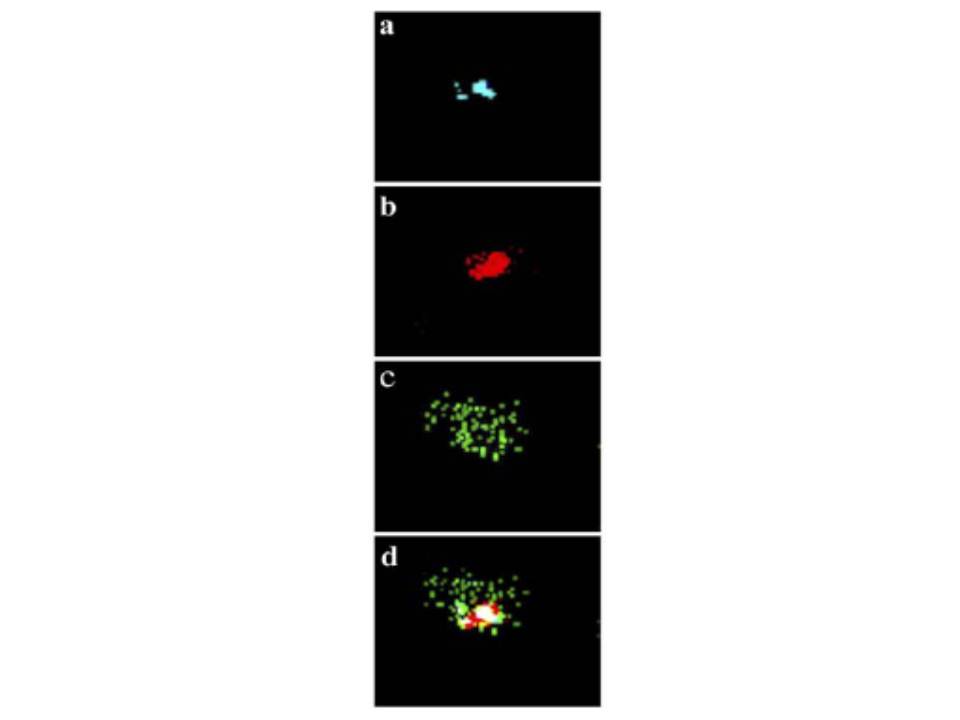
Confocal microscopy. Same field was observed with excitation wavelengths of 350 nm (DAPI), 579 nm (MitoTracker Red CMXRos) and 489 nm (Cy2). Blue, red and green fluorescence indicated the location of (a) nucleus (b) mitochondria and (c) PfCK, in Plasmodium falciparum, respectively. (d) Merged picture of above three individual pictures. The merged picture clearly suggested that PfCK is distributed throughout the cytoplasm (Fig. 6B, d) but not in the nucleus or mitochondria. Thus confocal studies further confirmed the cytosolic location of PfCK in P. falciparum.
Choubey V, Guha M, Maity P, Kumar S, Raghunandan R, Maulik PR, Mitra K, Halder UC, Bandyopadhyay U. Molecular characterization and localization of Plasmodium falciparum choline kinase. Biochim Biophys Acta. 2006 1760:1027-38.
