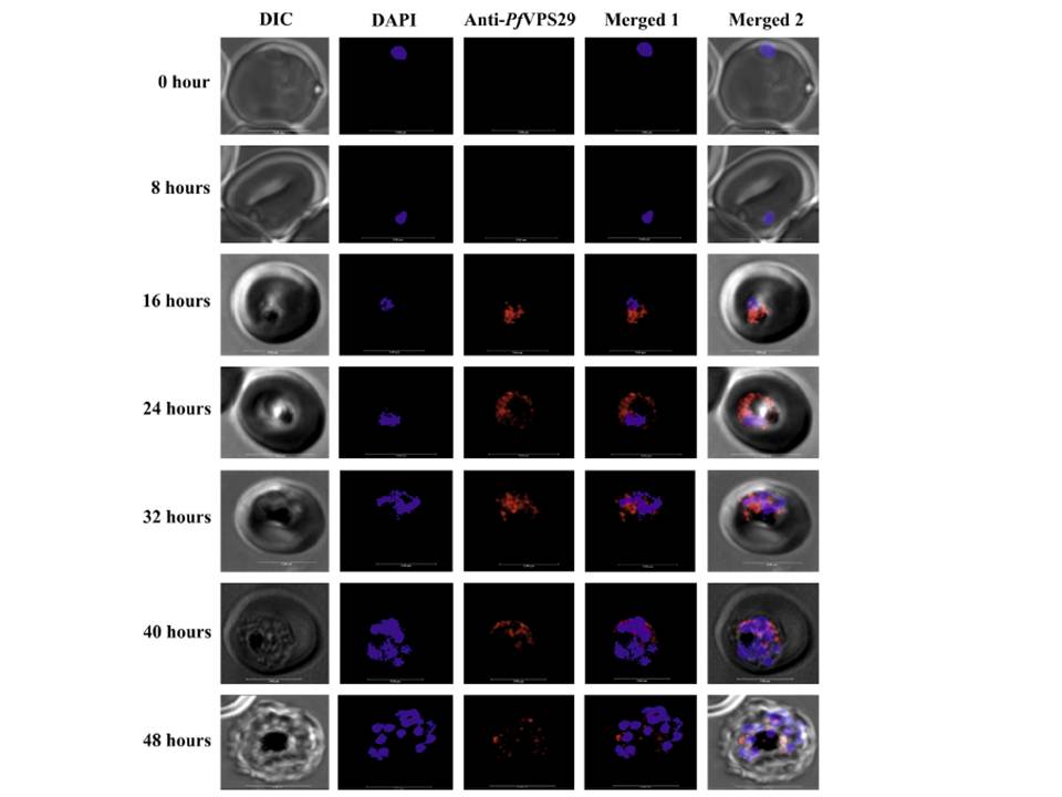
Localization of PfVPS29 in P. falciparum. Ring synchronized parasite (0 h) was grown in culture and at different time points of growth, iRBCs were processed and incubated with anti-PfVPS29 antibody followed by anti-rabbit Alexa fluor 647 (red fluorescence) and DAPI for nuclear staining (blue fluorescence). Each row is demonstrating a specific time point of growth from ring stage. Scale bar indicates 5 mm. PfVPS29 was located in the cytoplasm of the parasite. Although, PfVPS29 was clearly present from the trophozoite (16-32 h) up to the schizont (32-48 h) stage, its expression was observed maximally during the late trophozoite stage as evident from the fluorescent signal.
Iqbal MS, Siddiqui AA, Alam A, Goyal M, Banerjee C, Sarkar S, Mazumder S, De R, Nag S, Saha SJ, Bandyopadhyay U. Expression, purification and characterization of Plasmodium falciparum vacuolar protein sorting 29. Protein Expr Purif. 2015 Dec 12. [Epub ahead of print]
