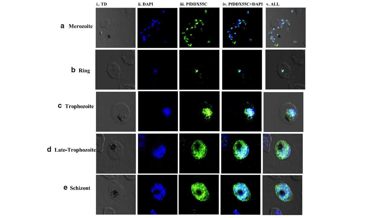
Localization of PfDDX55. Localization of PfDDX55 in different intraerythrocytic stages of P. falciparum by immunofluorescence staining and confocal microscopy. On a slide, fixed cells were immune stained with primary rabbit antibodies against PfDDX55C followed by Fluor isothiocyanate labeled secondary antibody and then counterstained with DAPI. In each panel, single confocal image of each stage is shown. Panel a–e show images stained with immune sera of PfDDX55C. a Merozoite stage, b ring stage, c trophozoite stage, d late trophozoite stage, and e schizont stage; In each panel, i- phase contrast (TD) image, ii- image of cell stained with DAPI (blue), iii- immuno-fluorescent stained cell (green), iv- superimposed image of ii and iii, v- merged image of panel i-iv. These data are representative of three separate experiments. The results show that it is expressed at all the intraerythrocytic stages. In merozoite, PfDDX55C is localized mainly in the cytoplasm (panels i-v). Whereas, in ring and early trophozoite stages, it is mainly localized in the nucleus (b, c, panels i-v). In later developmental stages like late trophozoite and schizont stages, it is localized throughout the parasite (both in nucleus and cytoplasm) (d, e).
Yasmin R, Kaur I, Tuteja R. Plasmodium falciparum DDX55 is a nucleocytoplasmic protein and a 3'-5' direction-specific DNA helicase. Protoplasma. 2020 [Epub ahead of print]