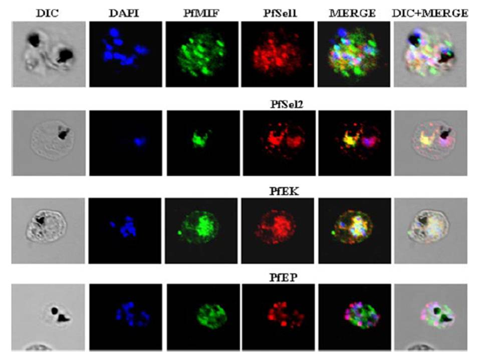
Indirect immunofluorescence assay and confocal microscopy to localize the selected excretory-secretory antigens by coimmunostaining of P. falciparum-infected erythrocytes. Air-dried infected erythrocytes were incubated with mouse antibodies specific to the selected proteins at 1:100 dilution (column 4; red). Rabbit aPfMIF antibody was used as a co-localization marker (column 3; green). The parasite nuclei were stained with 4’,6-diamidino-2-phenylindole (DAPI) (column 2; blue). Columns 1, 5, and 6 show a differential interference contrast (DIC) image, merged image, and differential interference contrast with merged image, respectively. Images showed punctate vesicle-like staining of the infected RBCs with antibodies to PfSEL1 (PFB0190c), PfSEL2 (Hrd3), PfEKand PfEP. PfEK also showing specific rimlike staining of the plasma membrane of the infected RBC. PfMIF is exported via the Maurer clefts to the extracellular medium after schizont rupture. A partial co-localization of PfMIF was seen with each of the four proteins, suggesting a distinct pathway for the release of these ESAs.
Singh M, Mukherjee P, Narayanasamy K, Arora R, Sen SD, Gupta S, Natarajan K, Malhotra P. Proteome analysis of Plasmodium falciparum extracellular secretory antigens at asexual blood stages reveals a cohort of proteins with possible roles in immune modulation and signaling. Mol Cell Proteomics. 2009 8(9):2102-18.
Other associated proteins
| PFID | Formal Annotation |
|---|---|
| PF3D7_1113100 | protein tyrosine phosphatase |
| PF3D7_1121300 | tyrosine kinase-like protein |
| PF3D7_1229400 | macrophage migration inhibitory factor |
