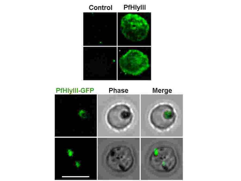
Upper panel: The localization of PfHly III was investigated by integrating the PfHly III-GFP fusion protein into the Dd2attb genome. The integration was done by transfecting the parasite with pLN-PfHly III and pINT. PCR analysis on transfected parasites using primers (GFP for, Hly III for and GFP rev) demonstrated the presence of pLN-PfHly III-GFP DNA. IFA with anti-His ab conjugated with alexa 488 showing Hly III coating the erythrocyte membrane. Lower panel: PfHly III-GFP localizes to the digestive vacuole of P. falciparum. Live fluorescence microscopy shows co-localization of 611 PfHly III-GFP with hemozoin. Scale bar, 7μm.
Moonah S, Sanders NG, Persichetti J, Sullivan DJ Jr. Erythrocyte lysis and Xenopus laevis oocyte rupture by recombinant Plasmodium falciparum hemolysin III. Eukaryot Cell. 2014 Aug 22. [Epub ahead of print]