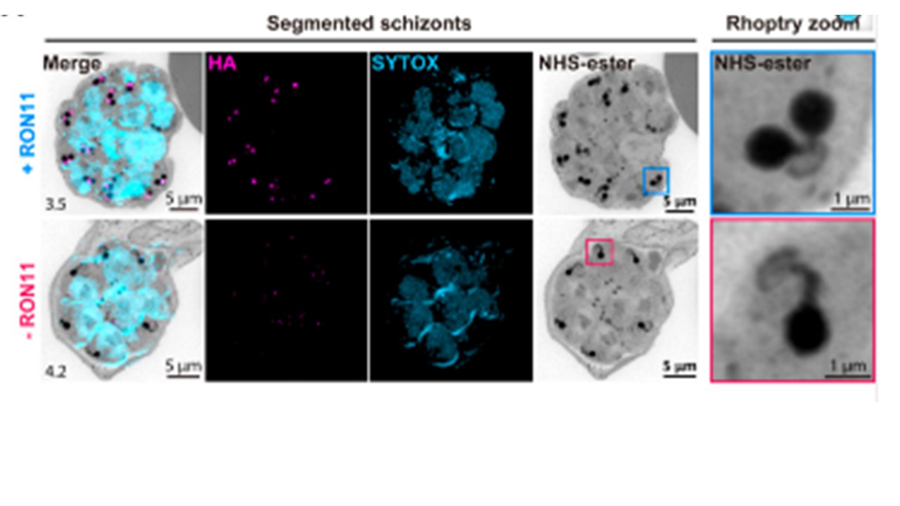
Representative U-ExM images of RON11apt schizonts showing the structure of rhoptries within fully developed merozoites in the presence or absence of 0.5 μm aTc. ML10-arrested schizonts were stained with NHS-Ester (grayscale), anti-HA (magenta), and the DNA dye SYTOX (cyan). Selected Z stack images were projected as a combined single image. Number on image = Z-axis thickness of projection in μm. (B) Quantification of the percentage of merozoites per schizont with single, dual, or multiple rhoptries in the presence or absence of 0.5 μm aTc. Samples were blinded and the number of rhoptries per merozoite within each schizont were counted. Merozoites were scored based on the number of nuclei observed (n = 4 biological replicates, 28 schizonts for +RON11 and 36 for -RON11; error bars = SEM; ****p < 0.0001 by unpaired two-tailed t test; the underlying data can be found in S1 Data). HA, hemagglutinin; NHS, N-hydroxysuccinimide; U-ExM, ultrastructure expansion microscopy. Anaguano D, Adewale-Fasoro O, Vick GW, Yanik S, Blauwkamp J, Fierro MA, Absalon S, Srinivasan P, Muralidharan V. Plasmodium RON11 triggers biogenesis of the merozoite rhoptry pair and is essential for erythrocyte invasion. PLoS Biol. 2024.