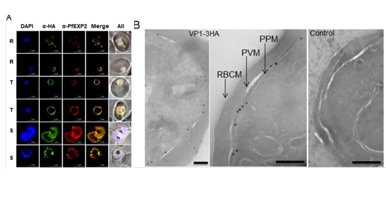
PfVP1 is mainly localized to the parasite plasma membrane. A, Immunofluorescence assay of Pf3D7VP2KO-VP1-3HAapt. DAPI stains the nuclei. Green, PfVP1-3HA. Red, PfEXP2. Note an ameboid ring stage parasite in the first row. R, ring. T, trophozoite. S, schizont. Scale bars, 5 μm.. B, Immunoelectron microscopy of Pf3D7VP2KO-VP1-3HAapt. RBCM, RBC membrane. PVM, parasitophorous vacuolar membrane. PPM, parasite plasma membrane. Scale bars, 200 nm. 3D7
wildtype served as control. C, Live cell imaging of Pf3D7VP2KO-VP1-mNeonGreenapt. M, merozoite. R, ring. T, trophozoite. S, schizont. Scale bar, 5 μm
Solebo O, Ling L, Nwankwo I, Zhou J, Fu TM, Ke H. Plasmodium falciparum utilizes pyrophosphate to fuel an essential proton pump in the ring stage and the transition to trophozoite stage. PLoS Pathog. 2023 19(12):e1011818. PMID: 38048362.
Other associated proteins
| PFID | Formal Annotation |
|---|---|
| PF3D7_1456800 | V-type H(+)-translocating pyrophosphatase, putative, VP1 |