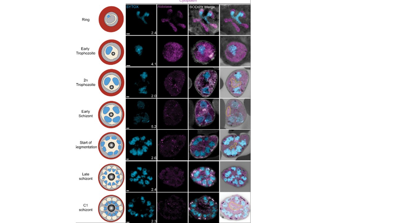
Cytoplasm staining during intraerythrocytic development.3D7 parasites were prepared by ultrastructural expansion microscopy (U-ExM), stained with N-hydroxysuccinimide (NHS) ester (grayscale), BODIPY TRc (white), SYTOX (cyan), and anti-aldolase (cytoplasm; magenta) antibodies and imaged by Airyscan microscopy across the asexual blood stage. Yellow line indicates likely position of food vacuole lacking hemozoin crystal. n = nucleus number. Images are maximum-intensity projections, number on image = Z-depth in µm of projection. Scale bars = 2 µm.