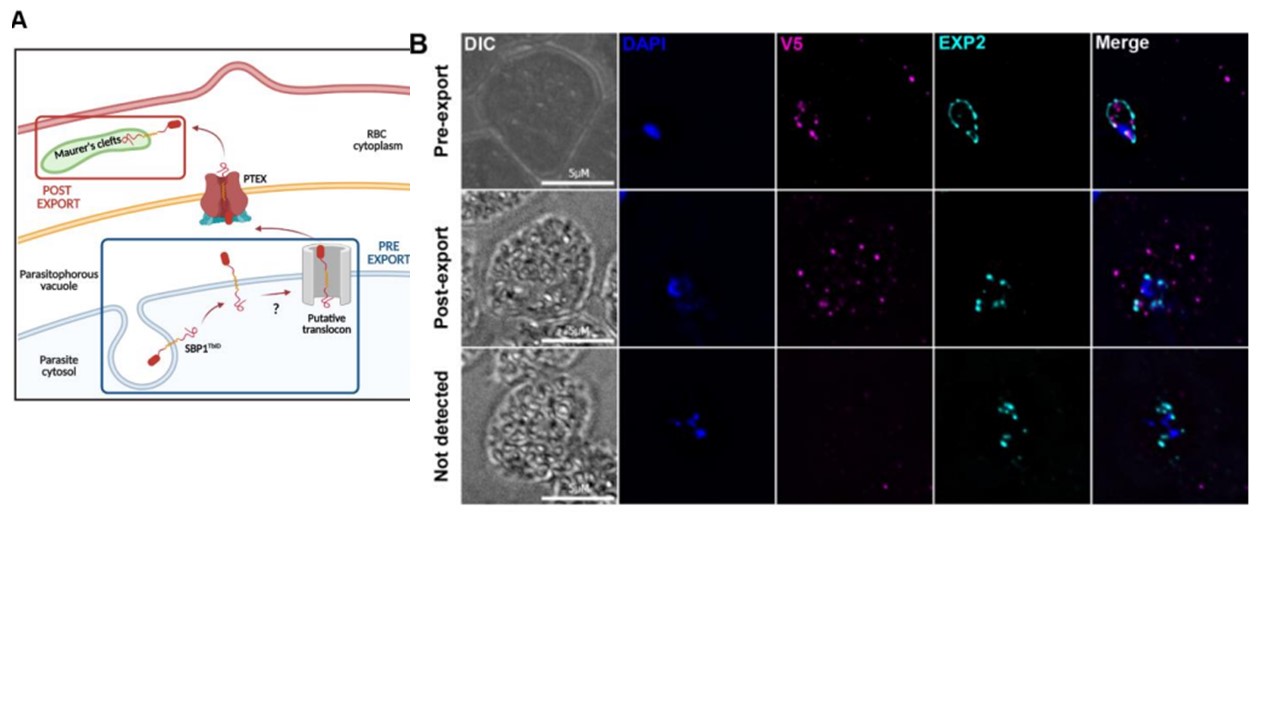
A) Schematic of the export of proteins in P. falciparum highlighting the locations where proteins biotinylated by SBP1TbID will be harvested. Created with BioRender.com. (B) IFA showing the different localizations of SBP1TbID during its export at early ring stages (3-5 hpi). Tightly synchronous SBP1TbID parasites were fixed with acetone at 3 h, 4h, and 5 h post-infection, and stained with specific antibodies. Images from left to right are phase-contrast, DAPI (nucleus, blue), anti-V5 (magenta), EXP2 (PV marker, cyan), and fluorescence merge. Z stack images were deconvolved and projected as a combined single image. Anaguano D, Dedkhad W, Brooks CF, Cobb DW, Muralidharan V. Time-resolved proximity biotinylation implicates a porin protein in export of transmembrane malaria parasite effectors. J Cell Sci. 2023 jcs.260506. PMID: 37772444.